Improve Physical and Mental Health during Aging with Mindfulness


Improve Physical and Mental Health during Aging with Mindfulness


By John M. de Castro, Ph.D.


“The healthier and more active one’s lifestyle, the more likely he or she will maintain cognitive performance over time. And meditation may be a key ingredient for ensuring brain health and maintaining good mental performance.“ – Grace Bullock


The aging process involves a systematic progressive decline in every system in the body, the brain included. The elderly frequently have problems with attention, thinking, and memory abilities, known as mild cognitive impairment. An encouraging new development is that mindfulness practices such as meditation training and mindful movement practices can significantly reduce these declines in cognitive ability. In addition, it has been found that

mindfulness practices reduce the deterioration of the brain that occurs with aging restraining the loss of neural tissue. Indeed, the brains of practitioners of meditation have been found to degenerate less with aging than non-practitioners.


In today’s Research News article “Long-Term Physical Exercise and Mindfulness Practice in an Aging Population.” (See summary below or view the full text of the study at: https://www.frontiersin.org/articles/10.3389/fpsyg.2020.00358/full?utm_source=F-AAE&utm_medium=EMLF&utm_campaign=MRK_1832518_a0P58000000G0YfEAK_Psycho_20220317_arts_A&utm_source=sfmc&utm_medium=email&utm_campaign=Article+Alerts+V4.1-Frontiers&utm_term=%%%3d+++++++REDIRECTTO(+++++CONCAT(%27http%3a%2f%2fjournal.frontiersin.org%2farticle%2f%27%2c+TreatAsContent(field(%40article%2c+%27DOI__c%27))%2c+%27%2ffull%3futm_source%3dF-AAE%26utm_medium%3dEMLF%26utm_campaign%3dMRK_%27%2c+TreatAsContent(JobID)%2c+%27_%27%2c+TreatAsContent(%40FieldId)%2c+%27_%27%2c+TreatAsContent(Substring(Replace(Field(%40field%2c+%27Name%27)%2c+%27+%27%2c+%27%27)%2c+0%2c+6))% ) Tang and colleagues compared older adults (average age of 64 years) who were either experienced (> 10 years) meditators or exercisers on physical, mental, immune, stress, and brain plasticity measures.


They report that the older adults who exercised had superior cardiovascular and respiratory fitness. But the older adults who meditated had superior physiological relaxation, quality of life, immune response, stress response, and brain plasticity. They conclude that the optimum results for older adults would be produced by combining meditation and exercise. Regardless, it is clear that meditation restrains the physical and mental deterioration with aging.


it’s heartening to know that age may not only bring wisdom or sore knees, but also more mindfulness.” – Jenn Director Knudsen 


CMCS – Center for Mindfulness and Contemplative Studies


This and other Contemplative Studies posts are also available on Twitter @MindfulResearch


Study Summary


Tang Y-Y, Fan Y, Lu Q, Tan L-H, Tang R, Kaplan RM, Pinho MC, Thomas BP, Chen K, Friston KJ and Reiman EM (2020) Long-Term Physical Exercise and Mindfulness Practice in an Aging Population. Front. Psychol. 11:358. doi: 10.3389/fpsyg.2020.00358


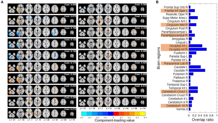
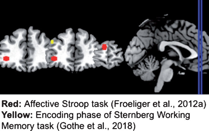
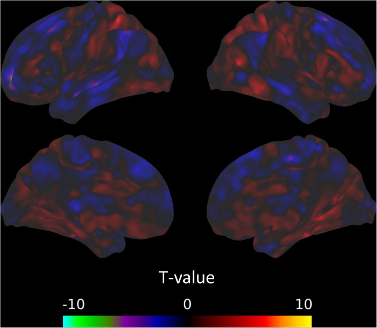
Previous studies have shown that physical exercise and mindfulness meditation can both lead to improvement in physical and mental health. However, it is unclear whether these two forms of training share the same underlying mechanisms. We compared two groups of older adults with 10 years of mindfulness meditation (integrative body-mind training, IBMT) or physical exercise (PE) experience to demonstrate their effects on brain, physiology and behavior. Healthy older adults were randomly selected from a large community health project and the groups were compared on measures of quality of life, autonomic activity (heart rate, heart rate variability, skin conductance response, respiratory amplitude/rate), immune function (secretory Immunoglobulin A, sIgA), stress hormone (cortisol) and brain imaging (resting state functional connectivity, structural differences). In comparison with PE, we found significantly higher ratings for the IBMT group on dimensions of life quality. Parasympathetic activity indexed by skin conductance response and high-frequency heart rate variability also showed more favorable outcomes in the IBMT group. However, the PE group showed lower basal heart rate and greater chest respiratory amplitude. Basal sIgA level was significantly higher and cortisol concentration was lower in the IBMT group. Lastly, the IBMT group had stronger brain connectivity between the dorsal anterior cingulate cortex (dACC) and the striatum at resting state, as well as greater volume of gray matter in the striatum. Our results indicate that mindfulness meditation and physical exercise function in part by different mechanisms, with PE increasing physical fitness and IBMT inducing plasticity in the central nervous systems. These findings suggest combining physical and mental training may achieve better health and quality of life results for an aging population.

https://www.frontiersin.org/articles/10.3389/fpsyg.2020.00358/full?utm_source=F-AAE&utm_medium=EMLF&utm_campaign=MRK_1832518_a0P58000000G0YfEAK_Psycho_20220317_arts_A&utm_source=sfmc&utm_medium=email&utm_campaign=Article+Alerts+V4.1-Frontiers&utm_term=%%%3d+++++++REDIRECTTO(+++++CONCAT(%27http%3a%2f%2fjournal.frontiersin.org%2farticle%2f%27%2c+TreatAsContent(field(%40article%2c+%27DOI__c%27))%2c+%27%2ffull%3futm_source%3dF-AAE%26utm_medium%3dEMLF%26utm_campaign%3dMRK_%27%2c+TreatAsContent(JobID)%2c+%27_%27%2c+TreatAsContent(%40FieldId)%2c+%27_%27%2c+TreatAsContent(Substring(Replace(Field(%40field%2c+%27Name%27)%2c+%27+%27%2c+%27%27)%2c+0%2c+6))%


Merry Mindful Christmas

3 Meditations to Get You in the Holiday Spirit

Merry Mindful Christmas


By John M. de Castro, Ph.D.


Christmas is not a time or season,
but a state of mind.
To cherish peace and goodwill,
to be plenteous in mercy,
is to have the real spirit of Christmas
.”

– Calvin Coolidge


Christmas is a Christian holiday celebrating the birth of Jesus. In fact, though, there is no historical records of the exact date of Jesus’ birth. December 25 was selected simply because it coincided with the very popular pagan celebrations of the Winter Solstice, allowing the Christians to coopt the celebration that they were unable to stop. Regardless of this history though Christmas has become a major celebration worldwide.


The Christian religions teach that this celebrates the birth of the savior, who came to earth to redeem us and save us from suffering. This event did not occur in a past. It occurred in the eternal present moment. We can also give birth to a savior. We can allow a personal mindfulness, a present moment awareness, to be born. If we do it can similarly be a savior. It can save us from much suffering. The suffering that we impose on ourselves.


The present moment contains only what is. Regrets and problems of the past don’t exist. Worries about the future don’t exist. If we accept it as it is and don’t constantly try to change it, it can bring us a relief of our worries and our woes. It can let us see the wonders of life and the happiness that is always present right here and right now. This surely would be a miraculous birth.


In our modern world Christmas has been commercialized and presents have become the focus. But as nice as it is to give and receive presents the joy is short-lived and can be followed almost immediately with disappointment and depression. In fact, this is the time of year when depression is rampant and the highest suicide rates of the year occurs.


The celebration would be much better if presence was the focus. The great sage Thich Nhat Hahn has said that the greatest gift that you can give another person is your presence. It is also true that the greatest gift that you can get is the presence of others. We’re not talking of simply being physically present, but being totally and deeply engaged with others; seeing, hearing, and interacting with them with full attention and caring. This produces a joy that lasts and warms from the inside. Indeed, the good feeling produced can last a lifetime.


In giving them our presence, we deeply observe and listen to them. We see all their non-verbal signals and attune our attention on what they are telling us. Without doing anything else this produces a very positive experience. How many times have we heard people tell us that they don’t feel noticed, that they don’t feel heard, and that they are not appreciated. Our presence is an antidote. We can satisfy these deep and often ignored deep needs. We can do this simply just by being there for them.


We may do this with the idea that we’re giving to others. But it quickly becomes obvious that this deeply affects us. The warmth radiates affecting all. Seeing the happiness in those we care about is deeply satisfying and a source of great happiness. So, in being present for others we bring them happiness and this, in turn, brings us happiness. This can produce an upward spiral of good feelings.


Celebrating a Christmas mindfully can amplify the good feelings and joy of the occasion. It can save us from our own self-produced suffering. It can help alleviate the suffering of others. It can bond us to others and them to us. It can make this holiday truly special.


So, merry mindful Christmas.


The way to a perfectly happy Christmas is to expect less and appreciate more!” – Marie Bloomfield


CMCS – Center for Mindfulness and Contemplative Studies


This and other Contemplative Studies posts are also available on Google+ https://plus.google.com/106784388191201299496/posts and on Twitter @MindfulResearch


Meditation Alters a Variety of Biological Mechanisms and Improves Mental Disorders

Meditation Alters a Variety of Biological Mechanisms and Improves Mental Disorders


By John M. de Castro, Ph.D.


Meditation-which come in many variations-has long been acknowledged as a tool to master the mind and cope with stress. Science is increasingly validating those claims, especially for depression, schizophrenia, anxiety, PTSD (post-traumatic stress disorder), and ADHD (attention deficit hyperactivity disorder).” – Mental Health America


Meditation training has been shown to improve health and well-being. It has also been found to be effective for a large array of medical and psychiatric conditions, either stand-alone or in combination with more traditional therapies. There are a number of ways that meditation practices produce these benefits, including changes to the brain and physiology. It is useful to review and summarize what has been discovered regarding the mechanisms by which meditation practice improves mental disorders.


In today’s Research News article “Biological mechanism study of meditation and its application in mental disorders.” (See summary below or view the full text of the study at: https://www.ncbi.nlm.nih.gov/pmc/articles/PMC7359050/) Shen and colleagues review and summarize the published scientific research studies on the mechanisms by which meditation practice improves mental disorders.


They report that the published research has found complex and widespread changes in the nervous system occur as a result of meditation. In the central nervous system these are relatively long lasting changes in the amount and connectivity of the brain tissue, termed neuroplastic changes, and these may underlie the beneficial changes in the meditators. In addition, meditation appears to alter the peripheral nervous system, in particular, the autonomic nervous system. Meditation increases parasympathetic activity that underlies vegetative functions and relaxation. This may be one mechanism by which meditation improves stress responses.


They further report that the published research found that meditation improves the functions of the immune and inflammatory systems. These effects also improve stress responses and fighting off disease. Hence, the effects of meditation on these biological process may underlie meditations ability to improve health. Since inflammatory responses often accompany mental illnesses, this may also be a mechanism by which meditation improved mental disease.


On a genetic, microbiological, level meditation has been found to alter the expression of genes that promote health. This may be the underlying reason that meditation improves the immune and inflammatory systems. Also, on the genetic level the research has found that meditation promotes the preservation of telomeres. These are the ends of the chromosomes that shorten throughout the lifetime and are thought to perhaps underlie cellular aging. This mechanism may underlie meditation’s ability to slow the aging process.


Meditation has been found through systematic controlled research to improve a wide array of mental illnesses. These include depression, including major depressive disorders, Post-traumatic stress disorder (PTSD), Attention Deficit Hyperactivity Disorder (ADHD), and Schizophrenia. In addition, meditation has been found to aid in recovery from substance abuse disorders and to help prevent relapse.


It is clear from the published scientific research that meditation alters a wide array of physiological processes and improves and improves an equally wide array of mental illnesses. It will be important in the future to link the two to begin to understand what physiological changes underlie which improvements in mental illness. Regardless it is clear that meditation has many beneficial effects that promote physical and mental well-being.


So, practice meditation to alter a variety of biological mechanisms and improve mental disorders.


Mindfulness exercises are valuable and useful for anyone, but most especially for people who are struggling with mental illness or addictions. “ – Sarah Levin


CMCS – Center for Mindfulness and Contemplative Studies


This and other Contemplative Studies posts are also available on Google+ https://plus.google.com/106784388191201299496/posts and on Twitter @MindfulResearch


Study Summary


Shen, H., Chen, M., & Cui, D. (2020). Biological mechanism study of meditation and its application in mental disorders. General psychiatry, 33(4), e100214. https://doi.org/10.1136/gpsych-2020-100214


Abstract

In recent years, research on meditation as an important alternative therapy has developed rapidly and been widely applied in clinical medicine. Mechanism studies of meditation have also developed progressively, showing that meditation has great impact on brain structure and function, and epigenetic and telomere regulation. In line with this, the application of meditation has gradually been expanded to mental illness, most often applied for major depressive disorders and substance-related and addictive disorders. The focus of this paper is to illustrate the biological mechanisms of meditation and its application in mental disorders.

Conclusions

Over the past two decades, meditation has been used in a great variety of fields to relieve stress, regulate emotions and promote physical and mental health. In recent years, the application of meditation in the psychiatric field has gradually received attention. It has become an adjunctive and alternative therapy for depression, PTSD and ADHD and has been carried out for the acute and remission stages of treatment for severe schizophrenia. Additionally, it can ameliorate emotional distress, craving and withdrawal symptoms in substance addiction. However, the current researchers adopt different meditation methods and diverse training durations, which leads to the inability to systematically evaluate which type of meditation is more beneficial to which populations or diseases, and to completely elucidate the biological mechanism of meditation. In the future, further targets for selective meditation subtypes along with prescribed training time, and randomised controlled studies with sufficient samples are required to determine the efficacy of meditation on the one hand, and simultaneously study the mechanisms behind meditation on the mind–body interaction, which can better display the positive function of meditation as an ancient physical and mental healing method in promoting human health.


https://www.ncbi.nlm.nih.gov/pmc/articles/PMC7359050/


Altered Brain Metabolism is Associated with Long-Term Yoga Practice

Altered Brain Metabolism is Associated with Long-Term Yoga Practice


By John M. de Castro, Ph.D.


the benefits of yoga are more encompassing than just the physical. And, thanks to modern technology and functional MRI scans, we’re now able to see how regular practice affects your brain.” – Emmy Lymn


The practice of yoga has many benefits for the individual’s physical and psychological health. Yoga has diverse effects because it is itself diverse having components of exercise, mindfulness meditation, and spirituality. So, yoga nourishes the body, mind, and spirit. As a result, yoga practice would be expected to produce physical changes. These include the relaxation response and stress relief. These should be obvious in the muscles, tendons and joints, but, less obvious in the nervous system. The nervous system changes in response to how it is used and how it is stimulated in a process called neuroplasticity. Highly used areas grow in size, metabolism, and connectivity. Mindfulness practices in general are known to produce these kinds of changes in the structure and activity of the brain.


In today’s Research News article “Long-term Ashtanga yoga practice decreases medial temporal and brainstem glucose metabolism in relation to years of experience.” (See summary below or view the full text of the study at: https://www.ncbi.nlm.nih.gov/pmc/articles/PMC7225240/), Aalst and colleagues recruited experienced adult yoga practitioners (at least 2 years of 3 times per week practice) and non-practitioners matched for age, gender, education, and physical activity levels. They had the experienced yoga practitioners perform 75 minutes of yoga while the control group practiced 75 minutes of aerobic exercise. Before and after they underwent a Positron Emission Tomography (PET) brain scan to determine changes in glucose metabolism (metabolic activity) in various brain regions.


They found that the experienced yoga practitioners at rest had significantly lower levels of activity in the hippocampus, parahippocampus, amygdala, insula, anterior midbrain, striatum (globus pallidus), and cerebellum compared to non-practitioners. After yoga practice there was a significant increase in activity in the cerebellum that wasn’t present for the non-practitioners after aerobic exercise. No significant differences in grey matter volume was observed.


The findings that the activity (brain metabolism) in the yoga practitioners while at rest is altered suggests that these are relatively permanent neuroplastic changes in the brain produced by long-term yoga practice. These changes are in areas that are known to be involved in mood and emotion regulation (limbic system, hippocampus, parahippocampus, amygdala), motor movements (cerebellum and striatum), and interoception and body awareness (Insula). These results are in line with the established ability of yoga practice to improve mood and emotion regulation, interoception and body awareness, and movement.


The findings are correlational and as such causation cannot be determined. But prior research has established that yoga training produces similar improvements in well-being and changes in the brain suggesting that these effects are caused by yoga practice. Yoga practice is a complex set of activities including postures, meditation, breathing practice, spirituality, and relaxation. It will remain for future research to determine which of these components or which combinations are responsible for which effects.


Yoga practitioners have different levels of brain activity at rest reflecting the psychological changes observed in yoga practitioners. The psychological changes suggest that the better emotional and physical well-being in yoga practitioners is due to neuroplastic changes in the brain produced by long-term yoga practice. These results support the recommendation of practicing yoga to improve physical and psychological well-being.


So, altered brain metabolism is associated with long-term yoga practice.


“The practice of yoga helps improve emotional regulation to reduce stress, anxiety and depression and that seems to improve brain functioning.” – Neha Gothe


CMCS – Center for Mindfulness and Contemplative Studies


This and other Contemplative Studies posts are also available on Google+ https://plus.google.com/106784388191201299496/posts and on Twitter @MindfulResearch


Study Summary


van Aalst, J., Ceccarini, J., Schramm, G., Van Weehaeghe, D., Rezaei, A., Demyttenaere, K., Sunaert, S., & Van Laere, K. (2020). Long-term Ashtanga yoga practice decreases medial temporal and brainstem glucose metabolism in relation to years of experience. EJNMMI research, 10(1), 50. https://doi.org/10.1186/s13550-020-00636-y


Abstract

Background

Yoga is increasingly popular worldwide with several physical and mental benefits, but the underlying neurobiology remains unclear. Whereas many studies have focused on pure meditational aspects, the triad of yoga includes meditation, postures, and breathing. We conducted a cross-sectional study comparing experienced yoga practitioners to yoga-naive healthy subjects using a multiparametric 2 × 2 design with simultaneous positron emission tomography/magnetic resonance (PET/MR) imaging.

Methods

18F-FDG PET, morphometric and diffusion tensor imaging, resting state fMRI, and MR spectroscopy were acquired in 10 experienced (4.8 ± 2.3 years of regular yoga experience) yoga practitioners and 15 matched controls in rest and after a single practice (yoga practice and physical exercise, respectively).

Results

In rest, decreased regional glucose metabolism in the medial temporal cortex, striatum, and brainstem was observed in yoga practitioners compared to controls (p < 0.0001), with a significant inverse correlation of resting parahippocampal and brainstem metabolism with years of regular yoga practice (ρ < − 0.63, p < 0.05). A single yoga practice resulted in significant hypermetabolism in the cerebellum (p < 0.0001). None of the MR measures differed, both at rest and after intervention.

Conclusions

Experienced yoga practitioners show regional long-term decreases in glucose metabolism related to years of practice. To elucidate a potential causality, a prospective longitudinal study in yoga-naive individuals is warranted.

https://www.ncbi.nlm.nih.gov/pmc/articles/PMC7225240/


Improve Emotion Processing by the Brain with Meditation

Improve Emotion Processing by the Brain with Meditation


By John M. de Castro, Ph.D.


“Alterations in key brain circuits associated with emotion regulation can be produced by mindfulness meditation.” – Richard Davidson


There has accumulated a large amount of research demonstrating that meditation practice has significant benefits for psychological, physical, and spiritual wellbeing. It has been shown to improve emotions and their regulation. Practitioners demonstrate more positive and less negative emotions and the ability to fully sense and experience emotions, while responding to them in appropriate and adaptive ways. In other words, mindful people are better able to experience yet control their responses to emotions. The ability of mindfulness training to improve emotion regulation is thought to be the basis for a wide variety of benefits that mindfulness provides to mental health and the treatment of mental illness especially depression and anxiety disorders.


One way that meditation practices may produce these benefits is by altering the brain. The nervous system is a dynamic entity, constantly changing and adapting to the environment. It will change size, activity, and connectivity in response to experience. These changes in the brain are called neuroplasticity. Over the last decade neuroscience has been studying the effects of contemplative practices on the brain and has identified neuroplastic changes in widespread areas. In other words, meditation practice appears to mold and change the brain, producing psychological, physical, and spiritual benefits.


In today’s Research News article “Meditation-induced neuroplastic changes of the prefrontal network are associated with reduced valence perception in older people.” (See summary below or view the full text of the study at: https://www.ncbi.nlm.nih.gov/pmc/articles/PMC7058252/), Chau and colleagues recruited adults 60 years of age or greater who had no meditation or relaxation training. They were randomly assigned to receive an 8-week program of 22 sessions of 1.5 hours each of either attention-based compassion meditation training or relaxation training. The participants were instructed to also practice at home daily. Before and after training they were measured for emotional valence (the difference between the magnitudes of positive and negative emotions) and arousal (overall magnitude of emotional responses relative to neutral) with an Emotional Processing task involving emotional ratings of positive neutral and negative pictures. They were also measured for attention with a Stroop task. In addition, they received a functional Magnetic Resonance Imaging (fMRI) brain scan.


They found that emotional valence and arousal significantly decreased after training for the meditation but not the relaxation group. This suggests that emotions were less extreme after meditation training. There were no significant differences with attention. The brain scans revealed that the meditation group had significant enlargements of the ventromedial prefrontal cortex, the inferior frontal sulcus, and the inferior frontal junction. Path analysis revealed the changes in the inferior frontal junction drove the changes in the ventromedial prefrontal cortex and the inferior frontal sulcus.


These results are interesting and demonstrate neuroplastic changes in the brains of the elderly produced by attention-based compassion meditation training but not relaxation training. These changes in the brains of the elderly are associated with decrease emotional reactivity. Indeed, the ventromedial prefrontal cortex has been shown to be involved in the inhibition of emotions. This suggests that the meditation training produced improved brain processing for the regulation of emotions in the elderly. Since the elderly often suffer from extremes of anxiety, depression, and loneliness, these meditation induced changes may improve the psychological health of the elderly.


So, improve emotion processing by the brain with meditation.


Meditation can help tame your emotions even if you’re not a mindful person.” – ScienceDaily


CMCS – Center for Mindfulness and Contemplative Studies


This and other Contemplative Studies posts are also available on Google+ https://plus.google.com/106784388191201299496/posts and on Twitter @MindfulResearch


Study Summary


Chau, B., Keuper, K., Lo, M., So, K. F., Chan, C., & Lee, T. (2018). Meditation-induced neuroplastic changes of the prefrontal network are associated with reduced valence perception in older people. Brain and Neuroscience Advances, 2, 2398212818771822. https://doi.org/10.1177/2398212818771822


Abstract

Background:

Neuroplastic underpinnings of meditation-induced changes in affective processing are largely unclear.

Methods:

We included healthy older participants in an active-controlled experiment. They were involved a meditation training or a control relaxation training of eight weeks. Associations between behavioral and neural morphometric changes induced by the training were examined.

Results:

The meditation group demonstrated a change in valence perception indexed by more neutral valence ratings of positive and negative affective images. These behavioral changes were associated with synchronous structural enlargements in a prefrontal network involving the ventromedial prefrontal cortex and the inferior frontal sulcus. In addition, these neuroplastic effects were modulated by the enlargement in the inferior frontal junction. In contrast, these prefrontal enlargements were absent in the active control group, which completed a relaxation training. Supported by a path analysis, we propose a model that describes how meditation may induce a series of prefrontal neuroplastic changes related to valence perception. These brain areas showing meditation-induced structural enlargements are reduced in older people with affective dysregulations.

Conclusion:

We demonstrated that a prefrontal network was enlarged after eight weeks of meditation training. Our findings yield translational insights in the endeavor to promote healthy aging by means of meditation.

https://www.ncbi.nlm.nih.gov/pmc/articles/PMC7058252/


Focused Meditation Changes Clustering of Brain Systems

Focused Meditation Changes Clustering of Brain Systems


By John M. de Castro, Ph.D.


meditation . . . appears to have an amazing variety of neurological benefits – from changes in grey matter volume to reduced activity in the “me” centers of the brain to enhanced connectivity between brain regions.” – Alice G. Walton


The nervous system is a dynamic entity, constantly changing and adapting to the environment. It will change size, activity, and connectivity in response to experience. These changes in the brain are called neuroplasticity.  Over the last decade neuroscience has been studying the effects of contemplative practices on the brain and has identified neuroplastic changes in widespread area. and have found that meditation practice appears to mold and change the brain, producing psychological, physical, and spiritual benefits. These brain changes with mindfulness practice are important and need to be further investigates.


Meditation practice results in a shift in mental processing. It produces a reduction of mind wandering and self-referential thinking and an increase in attention and higher-level thinking. The neural system that underlie mind wandering is termed the Default Mode Network (DMN) and consists in a set of brain structures including medial prefrontal cortex, posterior cingulate, lateral temporal cortex and the hippocampus. The neural system that underlies executive functions such as attention and higher-level thinking is termed the Fronto-Parietal Network (FPN). and includes the dorsolateral prefrontal cortex, posterior parietal cortex, and cingulate cortex.


There are a number of different types of meditation. Classically they’ve been characterized on a continuum with the degree and type of attentional focus. In focused attention meditation, the individual practices paying attention to a single meditation object. In today’s Research News article “Revealing Changes in Brain Functional Networks Caused by Focused-Attention Meditation Using Tucker3 Clustering.” (See summary below or view the full text of the study at: https://www.ncbi.nlm.nih.gov/pmc/articles/PMC6990115/), Miyoshi and colleagues examine the changes in the brain’s functional systems resulting from meditation practice. They recruited meditation naïve adults. They had their brains scanned with functional Magnetic Resonance Imaging (fMRI) during a 5-minute rest and a 5-minute breath-following (Focused) meditation.


They found in comparison to rest, during the brief focused meditation there was increased clustering in “eight brain regions, Frontal Inferior Operculum L, Occipital Inferior R, ParaHippocampal R, Cerebellum 10 R, Cingulum Middle R, Cerebellum Crus1 L, Occipital Inferior L, and Paracentral Lobule R increased through the meditation.” These are all regions involved in the Default Mode Network (DMN), the Somatosensory Network (SSN), and the Fronto-Parietal Network (FPN). The activity of these clusters best discriminated between the resting and focused meditative states.


These results make sense in that during a typical meditation there will be attentional focus, mind wandering, and return to attentional focus. The attentional focus is thought to involve the Fronto-Parietal Network (FPN). The mind wandering is thought to involve the Default Mode Network (DMN). Finally, returning from mind wandering to attentional focus is thought to involve Somatosensory Network (SSN). Hence the increased clustering in these systems seen in the focused meditative state would be expected given what is known of neural systems.


These results are from a very brief single focused meditation by meditation naïve participants. So, it does not reflect neuroplastic changes in the nervous system that would be expected in practiced meditators. Rather the results indicate the short term activation of clustered systems in the brain that if practiced over time would produce neuroplastic changes.


So, focused meditation changes clustering of brain systems.


long-term, active meditative practice decreases activity in the default network. This is the brain network associated with the brain at rest — just letting your mind wander with no particular goal in mind — and includes brain areas like the medial prefrontal cortex and the posterior cingulate cortex.” – Kayt Sukel


CMCS – Center for Mindfulness and Contemplative Studies


This and other Contemplative Studies posts are also available on Google+ https://plus.google.com/106784388191201299496/posts and on Twitter @MindfulResearch


Study Summary


Miyoshi, T., Tanioka, K., Yamamoto, S., Yadohisa, H., Hiroyasu, T., & Hiwa, S. (2020). Revealing Changes in Brain Functional Networks Caused by Focused-Attention Meditation Using Tucker3 Clustering. Frontiers in human neuroscience, 13, 473. doi:10.3389/fnhum.2019.00473


Abstract

This study examines the effects of focused-attention meditation on functional brain states in novice meditators. There are a number of feature metrics for functional brain states, such as functional connectivity, graph theoretical metrics, and amplitude of low frequency fluctuation (ALFF). It is necessary to choose appropriate metrics and also to specify the region of interests (ROIs) from a number of brain regions. Here, we use a Tucker3 clustering method, which simultaneously selects the feature vectors (graph theoretical metrics and fractional ALFF) and the ROIs that can discriminate between resting and meditative states based on the characteristics of the given data. In this study, breath-counting meditation, one of the most popular forms of focused-attention meditation, was used and brain activities during resting and meditation states were measured by functional magnetic resonance imaging. The results indicated that the clustering coefficients of the eight brain regions, Frontal Inferior Operculum L, Occipital Inferior R, ParaHippocampal R, Cerebellum 10 R, Cingulum Middle R, Cerebellum Crus1 L, Occipital Inferior L, and Paracentral Lobule R increased through the meditation. Our study also provided the framework of data-driven brain functional analysis and confirmed its effectiveness on analyzing neural basis of focused-attention meditation.

https://www.ncbi.nlm.nih.gov/pmc/articles/PMC6990115/


Yoga Practice Changes and Protects the Brain from Aging

Yoga Practice Changes and Protects the Brain from Aging


By John M. de Castro, Ph.D.


We can talk about anxiety, depression and blood pressure lowering in yoga, all of those are proven. But the biggest thing we see that results from yoga is that your quality of life will change for the better,” – Amy Wheeler


Human life is one of constant change. We revel in our increases in physical and mental capacities during development, but regret their decreases during aging. The aging process involves a systematic progressive decline in every system in the body, the brain included. Starting in the 20s there is a progressive decrease in the volume of the brain as we age. But the nervous system is a dynamic entity, constantly changing and adapting to the environment. It will change size, activity, and connectivity in response to experience. These changes in the brain are called neuroplasticity.


Over the last decade neuroscience has been studying the effects of contemplative practices on the brain and has identified neuroplastic changes in widespread area. and have found that meditation practice appears to mold and change the brain, producing psychological, physical, and spiritual benefits. In addition, they have been able to investigate various techniques that might slow the process of neurodegeneration that accompanies normal aging. They’ve found that mindfulness practices reduce the deterioration of the brain that occurs with aging restraining the loss of neural tissue. Indeed, the brains of practitioners of meditation and yoga have been found to degenerate less with aging than non-practitioners.


The evidence has been accumulating. It is reasonable to pause and summarize what has been learned. In today’s Research News article “Yoga Effects on Brain Health: A Systematic Review of the Current Literature.” (See summary below or view the full text of the study at: https://www.ncbi.nlm.nih.gov/pmc/articles/PMC6971819/), Gothe and colleagues review and summarize the published research studies of the effects of yoga practice on the brain. They found 11 published studies.


They report that the studies that compare the brains of yoga practitioners to non-practitioners and studies that trained participants in yoga have found increases in cortical volume and thickness particularly in the frontal cortex, hippocampus, anterior cingulate cortex and insula. They also found that yoga practice appears to increase the functional connectivity in a series of brain structures labelled as the default mode network. These changes are similar to those observed with other aerobic exercises. Importantly, the changes observed were mainly in the structures that are most affected by aging.


These findings from the currently available research studies suggest that yoga practice, like other aerobic exercises, can produce neuroplastic changes in the brain. These changes involve increases in size and function of areas that a typically seen to deteriorate with aging. This suggests that yoga practice can protect the brain from age-related deterioration. This would explain why yoga practice helps to prevent functional deterioration in the elderly.


These are important findings that suggest that yoga practice tends to protect or reverse age-related declines in the structure and functions of the nervous system. This could make for a healthier, happier aging process where the elderly retain cognitive abilities as they continue to age.


So, protect the brain from aging with yoga.


Using MRI scans, Villemure detected more gray matter—brain cells—in certain brain areas in people who regularly practiced yoga, as compared with control subjects.” – Stephani Sutherland


CMCS – Center for Mindfulness and Contemplative Studies


This and other Contemplative Studies posts are also available on Google+ https://plus.google.com/106784388191201299496/posts and on Twitter @MindfulResearch


Study Summary


Gothe, N. P., Khan, I., Hayes, J., Erlenbach, E., & Damoiseaux, J. S. (2019). Yoga Effects on Brain Health: A Systematic Review of the Current Literature. Brain plasticity (Amsterdam, Netherlands), 5(1), 105–122. doi:10.3233/BPL-190084


Abstract

Yoga is the most popular complementary health approach practiced by adults in the United States. It is an ancient mind and body practice with origins in Indian philosophy. Yoga combines physical postures, rhythmic breathing and meditative exercise to offer the practitioners a unique holistic mind-body experience. While the health benefits of physical exercise are well established, in recent years, the active attentional component of breathing and meditation practice has garnered interest among exercise neuroscientists. As the scientific evidence for the physical and mental health benefits of yoga continues to grow, this article aims to summarize the current knowledge of yoga practice and its documented positive effects for brain structure and function, as assessed with MRI, fMRI, and SPECT. We reviewed 11 studies examining the effects of yoga practice on the brain structures, function and cerebral blood flow. Collectively, the studies demonstrate a positive effect of yoga practice on the structure and/or function of the hippocampus, amygdala, prefrontal cortex, cingulate cortex and brain networks including the default mode network (DMN). The studies offer promising early evidence that behavioral interventions like yoga may hold promise to mitigate age-related and neurodegenerative declines as many of the regions identified are known to demonstrate significant age-related atrophy.

https://www.ncbi.nlm.nih.gov/pmc/articles/PMC6971819/


Reconfigure the Brain for Improved Executive Function with Meditation

Reconfigure the Brain for Improved Executive Function with Meditation


By John M. de Castro, Ph.D.


So, what’s the best way to build a better brain? Backed by 1000’s of studies, meditation is the neuroscientific community’s most proven way to upgrade the human brain.” – EOC Institute


The nervous system is a dynamic entity, constantly changing and adapting to the environment. It will change size, activity, and connectivity in response to experience. These changes in the brain are called neuroplasticity.  Over the last decade neuroscience has been studying the effects of contemplative practices on the brain and has identified neuroplastic changes in widespread area. and have found that meditation practice appears to mold and change the brain, producing psychological, physical, and spiritual benefits. These brain changes with mindfulness practice are important and need to be further investigates.


Meditation practice results in a shift in mental processing. It produces a reduction of mind wandering and self-referential thinking and an increase in attention and higher-level thinking. The neural system that underlie mind wandering is termed the Default Mode Network (DMN) and consists in a set of brain structures including medial prefrontal cortex, posterior cingulate, lateral temporal cortex and the hippocampus. The neural system that underlies executive functions such as attention and higher-level thinking is termed the Central Executive Network (CEN) and includes the dorsolateral prefrontal cortex, posterior parietal cortex, and cingulate cortex. Hence the shift in thought process may well be associated with changes in the relationship of these systems.


In today’s Research News article “From State-to-Trait Meditation: Reconfiguration of Central Executive and Default Mode Networks.” (See summary below or view the full text of the study at: https://www.ncbi.nlm.nih.gov/pmc/articles/PMC6893234/), Bauer and colleagues recruited experienced meditators and meditation naïve adults. Their brains were measured with functional Magnetic Resonance Imaging (fMRI) at rest (trait mindfulness) and while engaged in a brief meditation (state mindfulness).


They found that in comparison to the meditation naïve group during the resting state the experienced meditators had reduced activity and functional connectivity of the Default Mode Network (DMN) and reduced activity in the Central Executive Network (CEN) along with a stronger relationship between the activities of the DMN and CEN. These changes are indicative of the long-term changes in the neural systems produced by meditation and reflect the effects of trait mindfulness. During the meditation the experienced meditators had increased activity in the Central Executive Network (CEN) and increased functional connectivity with the Default Mode Network (DMN). These changes are indicative of the short-term changes in the neural systems produced by meditation and reflect the effects of state mindfulness.


These results suggest that long-term meditation practice alters the neural systems emphasizing reducing activation in both the mind wandering system (DMN) and the executive system (CEN) suggesting a reduction in thinking while at rest. This may be indicative of greater present moment awareness without evaluation or thought. The findings further suggest that long-term meditation practice alters the neural systems such that during meditation there is greater activity in the executive system (CEN) and greater influence of the CEN on the mind wandering system (DMN). This may be indicative of greater attention during meditation which suppresses mind wandering and self-referential thinking.


In general, it can be speculated that meditation practice alters the brain in ways that affect processing of information overall (trait), reducing thought and increasing awareness of the present moment environment. Meditation practice also alters the brain to increase the ability to attend during meditation and interrupt mind wandering. Hence, the brain activities reflect the subjective psychological changes seen in meditators.


So, reconfigure the brain for improved executive function with meditation.


“It seems the longer you do meditation, the better your brain will be at self-regulation. You don’t have to consume as much energy at rest and you can more easily get yourself into a more relaxed state.” – Bin He


CMCS – Center for Mindfulness and Contemplative Studies


This and other Contemplative Studies posts are e also available on Google+ https://plus.google.com/106784388191201299496/posts and on Twitter @MindfulResearch


Study Summary


Bauer, C., Whitfield-Gabrieli, S., Díaz, J. L., Pasaye, E. H., & Barrios, F. A. (2019). From State-to-Trait Meditation: Reconfiguration of Central Executive and Default Mode Networks. eNeuro, 6(6), ENEURO.0335-18.2019. doi:10.1523/ENEURO.0335-18.2019


Abstract

While brain default mode network (DMN) activation in human subjects has been associated with mind wandering, meditation practice has been found to suppress it and to increase psychological well-being. In addition to DMN activity reduction, experienced meditators (EMs) during meditation practice show an increased connectivity between the DMN and the central executive network (CEN). However, the gradual change between DMN and CEN configuration from pre-meditation, during meditation, and post-meditation is unknown. Here, we investigated the change in DMN and CEN configuration by means of brain activity and functional connectivity (FC) analyses in EMs across three back-to-back functional magnetic resonance imaging (fMRI) scans: pre-meditation baseline (trait), meditation (state), and post-meditation (state-to-trait). Pre-meditation baseline group comparison was also performed between EMs and healthy controls (HCs). Meditation trait was characterized by a significant reduction in activity and FC within DMN and increased anticorrelations between DMN and CEN. Conversely, meditation state and meditation state-to-trait periods showed increased activity and FC within the DMN and between DMN and CEN. However, the latter anticorrelations were only present in EMs with limited practice. The interactions between networks during these states by means of positive diametric activity (PDA) of the fractional amplitude of low-frequency fluctuations (fALFFs) defined as CEN fALFF¯¯¯¯¯¯¯¯¯¯¯¯¯¯¯¯¯¯¯¯ − DMN fALFF¯¯¯¯¯¯¯¯¯¯¯¯¯¯¯¯¯¯¯¯¯ revealed no trait differences but significant increases during meditation state that persisted in meditation state-to-trait. The gradual reconfiguration in DMN and CEN suggest a neural mechanism by which the CEN negatively regulates the DMN and is probably responsible for the long-term trait changes seen in meditators and reported psychological well-being.

https://www.ncbi.nlm.nih.gov/pmc/articles/PMC6893234/


Improve the Brain’s Attentional Networks with Mindfulness

Improve the Brain’s Attentional Networks with Mindfulness


By John M. de Castro, Ph.D.


MBSR and RR body scans both induced a common increased functional connectivity between the brain’s ventromedial prefrontal cortex, which plays a role in attention.” – GORAMA


The nervous system is a dynamic entity, constantly changing and adapting to the environment. It will change size, activity, and connectivity in response to experience. These changes in the brain are called neuroplasticity.  Over the last decade neuroscience has been studying the effects of contemplative practices on the brain and has identified neuroplastic changes in widespread area. and have found that meditation practice appears to mold and change the brain, producing psychological, physical, and spiritual benefits. These brain changes with mindfulness practice are important and need to be further investigates.


In today’s Research News article “Mindfulness-Based Stress Reduction-related changes in posterior cingulate resting brain connectivity.” (See summary below or view the full text of the study at: https://www.ncbi.nlm.nih.gov/pmc/articles/PMC6778831/), Kral and colleagues recruited healthy meditation-naïve adults and randomly assigned them to 8 weeks of Mindfulness-Based Stress Reduction (MBSR) program, 8 weeks of a Health Education Program, or to a wait-list control condition. The MBSR program consisted of 8 weekly group sessions involving meditation, yoga, body scan, and discussion. The participants were also encouraged to perform daily practice at home. The amount of home practice time was recorded. Before and after the 8-week intervention they were measured for emotional styles and participated in 14 days of experience sampling with 6 to 8 prompts per day via cellphone to indicate attention to task or mind wandering. They also underwent brain scanning with functional Magnetic Resonance Imaging (fMRI) before and after the intervention and 5.5 months later.


They found that in comparison to baseline and the health education and wait-list control groups, the participants who underwent the Mindfulness-Based Stress Reduction (MBSR) program had a significant increase in the functional connectivity between the posterior cingulate cortex and the dorsomedial prefrontal cortex. They also found that the higher the self-reported attention levels and the greater the number of days of practice the MBSR participants engaged in, the greater the increase in functional connectivity. The connectivity increases and the relationships with attention and practice were no longer significant at the 5.5-month follow-up. There were no significant changes in mind-wandering.


These results are interesting and suggest that participation in the Mindfulness-Based Stress Reduction (MBSR) program produces short-term changes in the brain’s system that underlies executive function and attention (the posterior cingulate cortex and the dorsomedial prefrontal cortex). The results further suggest that the amount of change in the brain system is associated with attentional changes and the amount of practice.


That mindfulness training in general and Mindfulness-Based Stress Reduction (MBSR) in particular improves attention and the neural systems underlying attention and executive function have been previously demonstrated. The present study demonstrates that these changes are related to the amounts of continuing practice suggesting the importance of practice outside of formal training sessions.


So, improve the brain’s attentional networks with mindfulness.


“mindfulness meditation decreases activity in the part of the brain that is in charge of mind-wandering and self-centeredness. Although we all struggle with taming that Monkey Mind, meditators are better at snapping out of it when the brain gets into a cycle of overthinking or negativity.” –  Jaime Carlo-Casellas


CMCS – Center for Mindfulness and Contemplative Studies


This and other Contemplative Studies posts are also available on Google+ https://plus.google.com/106784388191201299496/posts and on Twitter @MindfulResearch


Study Summary


Kral, T., Imhoff-Smith, T., Dean, D. C., Grupe, D., Adluru, N., Patsenko, E., … Davidson, R. J. (2019). Mindfulness-Based Stress Reduction-related changes in posterior cingulate resting brain connectivity. Social cognitive and affective neuroscience, 14(7), 777–787. doi:10.1093/scan/nsz050


Abstract

Mindfulness meditation training has been shown to increase resting-state functional connectivity between nodes of the frontoparietal executive control network (dorsolateral prefrontal cortex [DLPFC]) and the default mode network (posterior cingulate cortex [PCC]). We investigated whether these effects generalized to a Mindfulness-Based Stress Reduction (MBSR) course and tested for structural and behaviorally relevant consequences of change in connectivity. Healthy, meditation-naïve adults were randomized to either MBSR (N = 48), an active (N = 47) or waitlist (N = 45) control group. Participants completed behavioral testing, resting-state fMRI scans and diffusion tensor scans at pre-randomization (T1), post-intervention (T2) and ~5.5 months later (T3). We found increased T2–T1 PCC–DLPFC resting connectivity for MBSR relative to control groups. Although these effects did not persist through long-term follow-up (T3–T1), MBSR participants showed a significantly stronger relationship between days of practice (T1 to T3) and increased PCC–DLPFC resting connectivity than participants in the active control group. Increased PCC–DLPFC resting connectivity in MBSR participants was associated with increased microstructural connectivity of a white matter tract connecting these regions and increased self-reported attention. These data show that MBSR increases PCC–DLPFC resting connectivity, which is related to increased practice time, attention and structural connectivity.

https://www.ncbi.nlm.nih.gov/pmc/articles/PMC6778831/


Improve Tinnitus by Changing the Brain with Mindfulness

Improve Tinnitus by Changing the Brain with Mindfulness


By John M. de Castro, Ph.D.


The mindfulness approach is radically different from what most tinnitus sufferers have tried before, and it may not be right for everyone. We are confident, however, that the growing research base has demonstrated how it can offer an exciting new treatment to people who may have found that traditional treatment has not been able to help them yet.” – Liz Marks


Tinnitus is one of the most common symptoms to affect humanity. People with tinnitus live with a phantom noise that can range from a low hiss or ringing to a loud roar or squeal which can be present constantly or intermittently. It can have a significant impact on people’s ability to hear, concentrate, or even participate in everyday activities. Approximately 25 million to 50 million people in the United States experience it to some degree. Approximately 16 million people seek medical attention for their tinnitus, and for up to two million patients, debilitating tinnitus interferes with their daily lives.


There are a number of treatments for tinnitus including, counseling, sound therapy, drugs, and even brain stimulation. Unfortunately, none of these treatments is very effective. Mindfulness practices have been shown to be effective in treating Tinnitus. The nervous system is a dynamic entity, constantly changing and adapting to the environment. It will change size, activity, and connectivity in response to experience. These changes in the brain are called neuroplasticity. It is unknown how mindfulness practices may change the brain to improve tinnitus.


In today’s Research News article “Functional Brain Changes During Mindfulness-Based Cognitive Therapy Associated With Tinnitus Severity.” (See summary below or view the full text of the study at: https://www.ncbi.nlm.nih.gov/pmc/articles/PMC6667657/), Zimmerman and colleagues recruited adult participants in an 8-week Mindfulness-Based Cognitive Therapy (MBCT) program consisting of 2-hour weekly sessions and 40-60 minutes daily home practice. The MBCT program consists of mindfulness training and Cognitive Behavioral Therapy (CBT). During therapy the patient is trained to investigate and alter aberrant thought patterns underlying their reactions to tinnitus symptoms. The participants brains were scanned before and after the MBCT program, and at follow-up 8 weeks later with functional Magnetic resonance Imaging (fMRI) and were measured for tinnitus, anxiety, depression, and mindfulness.


They found that the MBCT program produced a significant reduction in tinnitus symptoms that were maintained at the 8-week follow-up. With the fMRI scans they found widespread changes in brain functional connectivity following the MBCT program. Significantly, they found a reduced connectivity between the amygdala and parietal cortex that was negatively correlated with the reduction in tinnitus symptoms. In other words, the greater the decrease in functional connectivity, the greater the reductions in tinnitus symptoms. It will require further research to determine how this connectivity change might be related to tinnitus symptoms.


The study demonstrated that the Mindfulness-Based Cognitive Therapy (MBCT) program reduces the symptoms of tinnitus in a lasting way. The brain scan results suggest that alterations of the functional connectivity of brain areas may underlie the symptom improvements. It will require considerably more research to determine the exact nature of the changes and their relationship to tinnitus. But the study is a good first start.


So, improve tinnitus by changing the brain with mindfulness.


“Mindfulness is a special kind of awareness: it . . . frees you to be more present in your immediate experience, so that you can wake up to the wonder of the one life you are given. Others have found that cultivating this practice has helped reduce the negative impact of tinnitus on their lives. The more open you can be to whatever you are experiencing at any moment, the more awake, alive, happy, and balanced you can be.” – Jennifer Gans

CMCS – Center for Mindfulness and Contemplative Studies


This and other Contemplative Studies posts are also available on Google+ https://plus.google.com/106784388191201299496/posts and on Twitter @MindfulResearch


Study Summary


Zimmerman, B., Finnegan, M., Paul, S., Schmidt, S., Tai, Y., Roth, K., … Husain, F. T. (2019). Functional Brain Changes During Mindfulness-Based Cognitive Therapy Associated With Tinnitus Severity. Frontiers in Neuroscience, 13, 747. doi:10.3389/fnins.2019.00747


Abstract

Mindfulness-based therapies have been introduced as a treatment option to reduce the psychological severity of tinnitus, a currently incurable chronic condition. This pilot study of twelve subjects with chronic tinnitus investigates the relationship between measures of both task-based and resting state functional magnetic resonance imaging (fMRI) and measures of tinnitus severity, assessed with the Tinnitus Functional Index (TFI). MRI was measured at three time points: before, after, and at follow-up of an 8-week long mindfulness-based cognitive therapy intervention. During the task-based fMRI with affective sounds, no significant changes were observed between sessions, nor was the activation to emotionally salient compared to neutral stimuli significantly predictive of TFI. Significant results were found using resting state fMRI. There were significant decreases in functional connectivity among the default mode network, cingulo-opercular network, and amygdala across the intervention, but no differences were seen in connectivity with seeds in the dorsal attention network (DAN) or fronto-parietal network and the rest of the brain. Further, only resting state connectivity between the brain and the amygdala, DAN, and fronto-parietal network significantly predicted TFI. These results point to a mostly differentiated landscape of functional brain measures related to tinnitus severity on one hand and mindfulness-based therapy on the other. However, overlapping results of decreased amygdala connectivity with parietal areas and the negative correlation between amygdala-parietal connectivity and TFI is suggestive of a brain imaging marker of successful treatment.

https://www.ncbi.nlm.nih.gov/pmc/articles/PMC6667657/