Mindfulness Improves Cognitive Function in Older Adults by Altering Brain Gene Expression

Mindfulness Improves Cognitive Function in Older Adults by Altering Brain Gene Expression


By John M. de Castro, Ph.D.


mindfulness may target inflammation, stress-related pathways, and neuroplasticity, thus reducing the risk of developing cerebrovascular disease and age-related neurodegeneration that could lead to the development of dementia.” – Ted Kheng Siang Ng


The aging process involves a systematic progressive decline in every system in the body, the brain included. The elderly frequently have problems with attention, thinking, and memory abilities, known as mild cognitive impairment. An encouraging new development is that mindfulness practices such as meditation training and mindful movement practices can significantly reduce these declines in cognitive ability. In addition, it has been found that

mindfulness practices reduce the deterioration of the brain that occurs with aging restraining the loss of neural tissue. Indeed, the brains of practitioners of meditation have been found to degenerate less with aging than non-practitioners. The mechanisms by which mindfulness affects the brain and reduces cognitive decline need to be investigated.


In today’s Research News article “Mindfulness intervention improves cognitive function in older adults by enhancing the level of miRNA-29c in neuron-derived extracellular vesicles.” (See summary below or view the full text of the study at: https://www.ncbi.nlm.nih.gov/pmc/articles/PMC8575875/ ) Hashizume and colleagues recruited healthy elderly adults aged 65 and over and administered either 4 weeks , 3 times per week for 60 minutes of Mindfulness-Based Stress Reduction (MBSR) or a no-treatment wait-list condition. They were measured before and after treatment for cognitive function including delayed recall, visuospatial/executive function, attention, abstraction, language, naming, and orientation tasks. They also had blood drawn and assayed for extracellular vesicles and mRNA in the vesicles.


They found that in comparison to baseline and the wait list control, after Mindfulness-Based Stress Reduction (MBSR) there were significant improvement in cognitive function including delayed recall, visuospatial/executive function, attention, naming, and orientation tasks. The blood assays revealed that in comparison to baseline and the wait list control, after MBSR there were significant reductions in miR-29c in the extracellular vesicles and decreased expression of the genes DNMT3A, DNMT3B, and BACE1 in in the extracellular vesicles. In another study with mice they found that injection into the brain ventricles of miR-29c prevented cognitive decline in the animals.


The results suggest that mindfulness training improves cognitive function in the elderly. These improvements in cognition have been previously observed by other researchers. The new findings in the present research are the changes in the extracellular vesicles found in the plasma. The expression of the mRNA miR-29c controls the gene expressions of DNMT3A, DNMT3B, and BACE1. These genes are associated with the loss of neurons in the brain. With aging there is a degeneration of the brain including losses of neurons. Reductions in the expression of the genes that tend to produce neuronal loss after Mindfulness-Based Stress Reduction (MBSR) suggests that the training protects the brain from the loss of neurons. This may represent the mechanism by which mindfulness training protects the brain in aging individual which results in improved cognitive function. It may be how mindfulness training stops cognitive decline in the elderly.


So, mindfulness improves cognitive function in older adults by altering brain gene expression.


an 8-week mindfulness-based training program improved cognition . . . in cognitively normal older adults, and that these improvements were associated with increased intrinsic connectivity within the default mode network.” – Gunes Sevinc


CMCS – Center for Mindfulness and Contemplative Studies


This and other Contemplative Studies posts are available on Twitter @MindfulResearch


Study Summary


Hashizume, S., Nakano, M., Kubota, K., Sato, S., Himuro, N., Kobayashi, E., Takaoka, A., & Fujimiya, M. (2021). Mindfulness intervention improves cognitive function in older adults by enhancing the level of miRNA-29c in neuron-derived extracellular vesicles. Scientific reports, 11(1), 21848. https://doi.org/10.1038/s41598-021-01318-y


Abstract

Although mindfulness-based stress reduction (MBSR) improves cognitive function, the mechanism is not clear. In this study, people aged 65 years and older were recruited from elderly communities in Chitose City, Japan, and assigned to a non-MBSR group or a MBSR group. Before and after the intervention, the Japanese version of the Montreal Cognitive Assessment (MoCA-J) was administered, and blood samples were collected. Then, neuron-derived extracellular vesicles (NDEVs) were isolated from blood samples, and microRNAs, as well as the target mRNAs, were evaluated in NDEVs. A linear mixed model analysis showed significant effects of the MBSR x time interaction on the MoCA-J scores, the expression of miRNA(miR)-29c, DNA methyltransferase 3 alpha (DNMT3A), and DNMT3B in NDEVs. These results indicate that MBSR can improve cognitive function by increasing the expression of miR-29c and decreasing the expression of DNMT3A, as well as DNMT3B, in neurons. It was also found that intracerebroventricular injection of miR-29c mimic into 5xFAD mice prevented cognitive decline, as well as neuronal loss in the subiculum area, by down-regulating Dnmt3a  and Dnmt3b  in the hippocampus. The present study suggests that MBSR can prevent neuronal loss and cognitive impairment by increasing the neuronal expression of miR-29c.

https://www.ncbi.nlm.nih.gov/pmc/articles/PMC8575875/


Improve Neuropsychological Disorders with Yoga

Improve Neuropsychological Disorders with Yoga


By John M. de Castro, Ph.D.


“yoga might be considered as an effective adjuvant for the patients with various neurological disorders including stroke, Parkinson’s disease, multiple sclerosis, epilepsy, Alzheimer’s disease, dementia, headache, myelopathy, neuropathies.” – A.Mooventhan


Mindfulness training and yoga practices have been shown to improve health and well-being in healthy individuals. They have also been found to be effective for a large array of medical and psychiatric conditions, either stand-alone or in combination with more traditional therapies. There has accumulated a large amount of research on the effectiveness of yoga practice for the treatment of a variety of physical and mental issues. Hence, it would be useful to summarize what has been learned.


In today’s Research News article “Therapeutic role of yoga in neuropsychological disorders.” (See summary below or view the full text of the study at: https://www.ncbi.nlm.nih.gov/pmc/articles/PMC8546763/ ) Nourollahimoghadam and colleagues review and summarize the published research regarding the effectiveness of yoga practice for the treatment of a variety of neuropsychological disorders.


They report that the published research found that yoga practice produced significant improvements in physical illnesses including migraine headaches, Alzheimer’s Disease, epilepsy, multiple sclerosis, Parkinson’s Disease, and neuropathy. Yoga practice also produced significant improvements in psychological well-being including anxiety, stress, depression, bipolar disorder, schizophrenia, somatoform disorders, obsessive-compulsive disorder, and burnout. They further report that yoga may produce its beneficial effects by altering the chemistry, electrical activity, structures, and connectivity within the brain.


Hence Yoga practice appears to have a myriad of positive physical and psychological benefits. The authors, however, point to weaknesses in the research including small sample sizes, short-term follow-up, confounding variables, and lack of appropriate controls. So, more and better controlled studies are needed to verify the benefits of yoga practice. Hence, the present state of knowledge supports the engagement in yoga practice to advance the physical and mental well-being of both ill and healthy individuals.


So, improve neuropsychological disorders with yoga.


Yoga can be a helpful practice of self-care for people with multiple sclerosis (MS) and other neurological conditions (such as stroke, traumatic brain injury, Parkinson’s disease, Lyme’s disease, Lou Gehrig’s disease).” – Mary Hilliker


CMCS – Center for Mindfulness and Contemplative Studies


This and other Contemplative Studies posts are also available on on Twitter @MindfulResearch


Study Summary


Nourollahimoghadam, E., Gorji, S., Gorji, A., & Khaleghi Ghadiri, M. (2021). Therapeutic role of yoga in neuropsychological disorders. World journal of psychiatry, 11(10), 754–773. https://doi.org/10.5498/wjp.v11.i10.754


Abstract

Yoga is considered a widely-used approach for health conservation and can be adopted as a treatment modality for a plethora of medical conditions, including neurological and psychological disorders. Hence, we reviewed relevant articles entailing various neurological and psychological disorders and gathered data on how yoga exerts positive impacts on patients with a diverse range of disorders, including its modulatory effects on brain bioelectrical activities, neurotransmitters, and synaptic plasticity. The role of yoga practice as an element of the treatment of several neuropsychological diseases was evaluated based on these findings.

Core Tip: A multitude of beneficial effects of yoga practice and the underlying mechanisms of action have been reported and point out its role as an influential element in the integrative therapy of various neuropsychological disorders. In the planning of further investigations, studies should be designed to achieve more accuracy and precision in the heterogeneous field of yoga practices and potential fields of application.

https://www.ncbi.nlm.nih.gov/pmc/articles/PMC8546763/


Hallucinogenic Drugs may Effectively Treat Mental Illness

Hallucinogenic Drugs may Effectively Treat Mental Illness


By John M. de Castro, Ph.D.


Combined with psychotherapy, some psychedelic drugs like MDMA, psilocybin and ayahuasca may improve symptoms of anxiety, depression and post-traumatic stress disorder,” – Cristina L. Magalhaes


Psychedelic substances such as peyote, mescaline, LSD, Bufotoxin, ayahuasca and psilocybin 

have been used almost since the beginning of recorded history to alter consciousness and produce spiritually meaningful experiences. More recently hallucinogenic drugs such as MDMA (Ecstasy) and Ketamine have been similarly used. People find the experiences produced by these substances extremely pleasant. eye opening, and even transformative. They often report that the experiences changed them forever. Psychedelics and hallucinogens have also been found to be clinically useful as they markedly improve mood, increase energy and enthusiasm and greatly improve clinical depression. The research on the effectiveness of these drugs on mood and mental illness is accumulating. So, it makes sense to step back and summarize what has been learned.


In today’s Research News article “.” (See summary below or view the full text of the study at: https://www.ncbi.nlm.nih.gov/pmc/articles/PMC7880300/ ) De Gregorio and colleagues review and summarize the published research on the brain mechanisms of hallucinogenic drug actions and their effectiveness as treatments for mental illness.


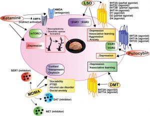
They report that the different drugs have very different effects on the nervous system although most interact with serotonin receptors. The nervous systems effects appear to alter sensory integration and associations with these sensations resulting in altered experiences.


They also report that the published research suggests that psilocybin may be useful in treating anxiety, depression, alcohol abuse, and obsessive-compulsive disorder (OCD), LSD may relieve anxiety and depression, that Ketamine may improve major depressive disorder, and MDMA (Ecstasy) may help in the treatment of post-traumatic stress disorder. Hence, psychedelic and hallucinogenic drugs may be effective in treating mental illness. It needs to be kept in mind that these drugs have powerful effects so they must be administered in controlled environments by trained practitioners.


So, hallucinogenic drugs may effectively treat mental illness.


Most powerful substances that we know of, that have powerful effects on the central nervous system, are like any powerful tool, They can have dangerous effects, or beneficial effects, if judiciously used in a context where the dangers are known and mechanisms are in place to address them.” – Matthew Johnson


CMCS – Center for Mindfulness and Contemplative Studies


This and other Contemplative Studies posts are also available on Google+ https://plus.google.com/106784388191201299496/posts and on Twitter @MindfulResearch


Study Summary


De Gregorio, D., Aguilar-Valles, A., Preller, K. H., Heifets, B. D., Hibicke, M., Mitchell, J., & Gobbi, G. (2021). Hallucinogens in Mental Health: Preclinical and Clinical Studies on LSD, Psilocybin, MDMA, and Ketamine. The Journal of neuroscience : the official journal of the Society for Neuroscience, 41(5), 891–900. https://doi.org/10.1523/JNEUROSCI.1659-20.2020


Abstract

A revamped interest in the study of hallucinogens has recently emerged, especially with regard to their potential application in the treatment of psychiatric disorders. In the last decade, a plethora of preclinical and clinical studies have confirmed the efficacy of ketamine in the treatment of depression. More recently, emerging evidence has pointed out the potential therapeutic properties of psilocybin and LSD, as well as their ability to modulate functional brain connectivity. Moreover, MDMA, a compound belonging to the family of entactogens, has been demonstrated to be useful to treat post-traumatic stress disorders. In this review, the pharmacology of hallucinogenic compounds is summarized by underscoring the differences between psychedelic and nonpsychedelic hallucinogens as well as entactogens, and their behavioral effects in both animals and humans are described. Together, these data substantiate the potentials of these compounds in treating mental diseases.

https://www.ncbi.nlm.nih.gov/pmc/articles/PMC7880300/


Hot Yoga is Superior in Improving Cardiovascular Health

Hot Yoga is Superior in Improving Cardiovascular Health


By John M. de Castro, Ph.D.


“It might be hard, but hot yoga can help improve strength, flexibility and balance, while helping decrease your risk of conditions such as diabetes and heart disease.” – Lydia Stephens


Cardiovascular disease is the number one killer, claiming more lives than all forms of cancer combined. “Heart disease is the leading cause of death for both men and women. About 610,000 people die of heart disease in the United States every year–that’s 1 in every 4 deaths. Every year about 735,000 Americans have a heart attack.” (Centers for Disease Control). A myriad of treatments has been developed for heart disease including a variety of surgical procedures and medications. But the safest effective treatments are lifestyle changes. These include quitting smoking, weight reduction, improved diet, physical activity, and reducing stresses. Other safe and effective treatments are contemplative practices, such as meditation, tai chi, and yoga, have also been shown to be helpful for heart health. These practices have also been shown to be helpful for producing the kinds of lifestyle changes needed such as smoking cessationweight reduction, and stress reduction.


Yoga is a mindfulness practice and exercise that has been shown to improve physical well-being and cardiovascular health. Hot Yoga is somewhat unique yoga practice as it is practiced in a heated environment (105°F, 40.6°C, 40% humidity). The hot environment is thought to soften the muscles making them more pliable and loosen the joints making them more flexible allowing the practitioner to go deeper into poses. The sweating that occurs is thought to help remove toxins and impurities. It is not known if hot yoga is better in improving cardiovascular health than regular yoga.


In today’s Research News article “Cardiovascular, Cellular, and Neural Adaptations to Hot Yoga versus Normal-Temperature Yoga.” (See summary below or view the full text of the study at: https://www.ncbi.nlm.nih.gov/pmc/articles/PMC8191229/ ) Bourbeau and colleagues recruited adults and randomly assigned them to either practice normal yoga or hot yoga for 75 minutes 3 times per week for 4 weeks. Both normal and hot yoga sessions followed an identical taped standardized yoga program. The participants were measured before and after training for body size, maximal aerobic capacity (VO2Max), heat tolerance, and perceived stress. In addition, blood was drawn and assayed for adrenocorticotropic hormone, brain-derived neurotrophic factor, and heat shock protein 70.


They found that in comparison to baseline the hot yoga group had significant increases in maximal aerobic capacity (VO2Max) and heat shock protein 70, while the normal yoga group did not. Both yoga groups had significant increases in brain-derived neurotrophic factor. Neither yoga practices significantly altered plasma adrenocorticotropic hormone, perceived stress, or heat tolerance.


These results suggest that practicing yoga in a hot environment increases its ability to improve cardiovascular fitness (VO2Max) and cellular heat tolerance (Heat shock protein 70). Yoga in either form increase brain development (Brain-derived neurotrophic factor). This suggests for adult patients at risk for or having cardiovascular problems who can also tolerate high temperatures that hot yoga may be an effective treatment to improve cardiovascular function. The results also show as has been shown in multiple previous studies that yoga practice improve the physical well-being of the participants.


So, hot yoga is superior in improving cardiovascular health.


hot yoga promises a number of compelling health benefits, alleging to cure everything from back pain to high blood pressure to anxiety.” – Anna Dickens


CMCS – Center for Mindfulness and Contemplative Studies


This and other Contemplative Studies posts are also available on Google+ https://plus.google.com/106784388191201299496/posts and on Twitter @MindfulResearch


Study Summary


Bourbeau, K. C., Moriarty, T. A., Bellovary, B. N., Bellissimo, G. F., Ducharme, J. B., Haeny, T. J., & Zuhl, M. N. (2021). Cardiovascular, Cellular, and Neural Adaptations to Hot Yoga versus Normal-Temperature Yoga. International Journal of Yoga, 14(2), 115–126. https://doi.org/10.4103/ijoy.IJOY_134_20


Abstract

Context:

Chronic heat exposure promotes cardiovascular and cellular adaptations, improving an organism’s ability to tolerate subsequent stressors. Heat exposure may also promote neural adaptations and alter the neural–hormonal stress response. Hot-temperature yoga (HY) combines mind–body exercise with heat exposure. The added heat component in HY may induce cardiovascular and cellular changes, along with neural benefits and modulation of stress hormones.

Aims:

The purpose of the present study is to compare the cardiovascular, cellular heat shock protein 70 (HSP70), neural, and hormonal adaptations of HY versus normal-temperature yoga (NY).

Settings and Design:

Twenty-two subjects (males = 11 and females = 11, 26 ± 6 years) completed 4 weeks of NY (n = 11) or HY (n = 11, 41°C, 40% humidity). Yoga sessions were performed 3 times/week following a modified Bikram protocol.

Subjects and Methods:

Pre- and posttesting included (1) hemodynamic measures during a heat tolerance test and maximal aerobic fitness test; (2) neural and hormonal adaptations using serum brain-derived neurotrophic factor (BDNF) and adrenocorticotropic hormone (ACTH), along with a mental stress questionnaire; and (3) cellular adaptations (HSP70) in peripheral blood mononuclear cells (PBMCs).

Statistical Analysis:

Within- and between-group Student’s t-test analyses were conducted to compare pre- and post-VO2 max, perceived stress, BDNF, HSP70, and ACTH in HY and NY groups.

Results:

Maximal aerobic fitness increased in the HY group only. No evidence of heat acclimation or change in mental stress was observed. Serum BDNF significantly increased in yoga groups combined. Analysis of HSP70 suggested higher expression of HSP70 in the HY group only.

Conclusions:

Twelve sessions of HY promoted cardiovascular fitness and cellular thermotolerance adaptations. Serum BDNF increased in response to yoga (NY + HY) and appeared to not be temperature dependent.

https://www.ncbi.nlm.nih.gov/pmc/articles/PMC8191229/


Yoga Practice Improves Glycemic Control in Type 2 Diabetes

Yoga Practice Improves Glycemic Control in Type 2 Diabetes


By John M. de Castro, Ph.D.


“Yoga can do more than just relax your body in mind — especially if you’re living with diabetes. Certain poses may help lower blood pressure and blood sugar levels while also improving circulation, leading many experts to recommend yoga for diabetes management.” – Healthline


Diabetes is a major health issue. It is estimated that 30 million people in the United States and nearly 600 million people worldwide have diabetes and the numbers are growing. Type II Diabetes results from a resistance of tissues, especially fat tissues, to the ability of insulin to promote the uptake of glucose from the blood. As a result, blood sugar levels rise producing hyperglycemia. Diabetes is heavily associated with other diseases such as cardiovascular disease, heart attacks, stroke, blindness, kidney disease, and circulatory problems leading to amputations. As a result, diabetes doubles the risk of death of any cause compared to individuals of the same age without diabetes.


Type 2 diabetes is a common and increasingly prevalent illness that is largely preventable. One of the reasons for the increasing incidence of Type 2 Diabetes is its association with overweight and obesity which is becoming epidemic in the industrialized world. A leading cause of this is a sedentary life style. Unlike Type I Diabetes, Type II does not require insulin injections. Instead, the treatment and prevention of Type 2 Diabetes focuses on diet, exercise, and weight control. Recently, mindfulness practices have been shown to be helpful in managing diabetes. A mindfulness practice that combines mindfulness with exercise is yoga and it has been shown to be helpful in the treatment of Type II Diabetes.


In today’s Research News article “Effectiveness of Yoga-based Exercise Program Compared to Usual Care, in Improving HbA1c in Individuals with Type 2 Diabetes: A Randomized Control Trial.” (See summary below or view the full text of the study at: https://www.ncbi.nlm.nih.gov/pmc/articles/PMC7735507/ ) Gupta and colleagues recruited  patients over 30 years of age with Type 2 Diabetes. They were randomly assigned to receive either a 4-month program of diet and walking exercise or a Yoga-based Exercise Program. The yoga program consisted of meditation, breathing exercises, postures, and stretching. The participants had blood drawn before and after training and assayed for HbA1c, blood fats, and blood glucose.


They found that in comparison to baseline and the diet and walking group, the yoga group had a significantly greater reduction in HbA1c. Glycated hemoglobin, HbA1c, is a measure that reflect the control of blood glucose levels over a long period of time. Low HbA1c levels is a sign of good blood glucose control which is essential to the control of the effects of Type 2 Diabetes on the body. So, the results signal that yoga practice results in better glucose control in these patients than a diet and walking program. This suggests that yoga would promote better health in patients with Type 2 Diabetes.


So, yoga practice improves glycemic control in Type 2 Diabetes.


Stress elevates blood sugar, which can lead to more diabetes complications. Yoga helps us center ourselves, and centering calms us and can help keep blood sugar levels balanced.” – Janet Zappe


CMCS – Center for Mindfulness and Contemplative Studies


This and other Contemplative Studies posts are also available on Google+ https://plus.google.com/106784388191201299496/posts and on Twitter @MindfulResearch


Study Summary


Gupta, U., Gupta, Y., Jose, D., Mani, K., Jyotsna, V. P., Sharma, G., & Tandon, N. (2020). Effectiveness of Yoga-based Exercise Program Compared to Usual Care, in Improving HbA1c in Individuals with Type 2 Diabetes: A Randomized Control Trial. International journal of yoga, 13(3), 233–238. https://doi.org/10.4103/ijoy.IJOY_33_20


Abstract

Background:

This study was designed to evaluate the effectiveness of a yoga-based exercise program (YBEP) in improving glycemic control in people with type 2 diabetes mellitus.

Materials and Methods:

Patients on stable oral glucose-lowering agents for at least 3 months and HbA1c 7.5%–10% were randomized in 1:1 ratio. The primary outcome measure was the difference of change in mean HbA1c between groups.

Results:

The participants (n = 81) had mean (±standard deviation) age of 50.6 (±8.5) years and HbA1c of 8.5 ± 0.7% (68.97 ± 7.42 mmol/mol). The follow-up data were available in 96% (78/81) of participants. Of 40 participants, 25 (62.5%) attended ≥75% (≥10 out of 13) of the sessions in YBEP. On the intention to treat analysis, a favorable reduction (0.21% 95% confidence interval [−0.34, 0.75], P = 0.454) in HbA1c was seen in YBEP group as compared to usual care. The reduction in HbA1c by ≥0.5% was observed in 44.7% of participants in YBEP as compared to 37.5% in usual care arm, respectively. Those who attended ≥75% of the sessions had better HbA1c reduction of 0.3% in comparison to 0.1% reduction seen in those who attended <75% of the sessions.

Conclusions:

YBEP demonstrated a clinically relevant HbA1c reduction compared to usual care in participants who had attended at least 75% of the yoga sessions. The reduction in HbA1c by >0.5% in 44.7% in the yoga group, suggests, that it can be prescribed as an exercise to individuals who are unable to walk either due to limited joint mobility, adverse weather conditions, lack of space for walking, cultural or religious prohibitions for women for outdoor physical activity, and so on.

https://www.ncbi.nlm.nih.gov/pmc/articles/PMC7735507/


Increase Brain-Derived Neurotrophic Factor (BDNF) with Mindfulness

Increase Brain-Derived Neurotrophic Factor (BDNF) with Mindfulness


By John M. de Castro, Ph.D.


“BDNF is a key neurotrophin that promotes development, survival and plasticity of neurons in the central and peripheral nervous systems.” – B. Rael Cahn


The nervous system is a dynamic entity, constantly changing and adapting to the environment. It will change size, activity, and connectivity in response to experience. These changes in the brain are called neuroplasticity.  Over the last decade neuroscience has been studying the effects of contemplative practices on the brain and has identified neuroplastic changes in widespread area. and have found that meditation practice appears to mold and change the brain, producing psychological, physical, and spiritual benefits. These brain changes with mindfulness practice are important and need to be further investigates.


Brain-Derived Neurotrophic Factor (BDNF) is a key molecule circulating in the blood that promotes neural, neuronal survival and regeneration which produces neuroplasticity. There have been a number of studies performed on the ability of Mindfulness practices to affect the levels of BDNF found in the blood. It makes sense, then to review what has been learned.


In today’s Research News article “The Effect of Mindfulness-Based Intervention on Brain-Derived Neurotrophic Factor (BDNF): A Systematic Review and Meta-Analysis of Controlled Trials.” (See summary below or view the full text of the study at: https://www.frontiersin.org/articles/10.3389/fpsyg.2020.02209/full?utm_source=F-AAE&utm_medium=EMLF&utm_campaign=MRK_1437459_69_Psycho_20200922_arts_A ) Gomutbutra and colleagues review, summarize, and perform a meta-analysis of the effects of mindfulness practices on the circulating levels of Brain-Derived Neurotrophic Factor (BDNF). They identified 11 published randomized controlled trials.


They report that the published research studies found that mindfulness training either by meditation or yoga practice produced a significant increase in circulating levels of Brain-Derived Neurotrophic Factor (BDNF). Hence the published research suggests that meditation and yoga practice produce increased levels of BDNF in the blood. Since BDNF promotes neuroplasticity, this may be the mechanism by which mindfulness practices change the brain and thereby improve health and well-being. Of course, much more research is needed but this is a promising potential mechanism whereby mindfulness produces its benefits.


So, Increase Brain-Derived Neurotrophic Factor (BDNF) with Mindfulness.


mindfulness meditation and mind-body exercise (e.g. yoga and tai chi) increase circulating BDNF concentrations in healthy and diseased individuals.” – Tongjian You


CMCS – Center for Mindfulness and Contemplative Studies


This and other Contemplative Studies posts are also available on Google+ https://plus.google.com/106784388191201299496/posts and on Twitter @MindfulResearch


Study Summary


Gomutbutra P, Yingchankul N, Chattipakorn N, Chattipakorn S and Srisurapanont M (2020) The Effect of Mindfulness-Based Intervention on Brain-Derived Neurotrophic Factor (BDNF): A Systematic Review and Meta-Analysis of Controlled Trials. Front. Psychol. 11:2209. doi: 10.3389/fpsyg.2020.02209


Background: This systematic review aims to answer three questions. First, how much do mindfulness-based interventions (MBIs) affect peripheral brain-derived neurotrophic factor (BDNF)? Second, do mindfulness exercise–based interventions (exercise-MBIs) and mindfulness meditation–based interventions (meditation-MBIs) affect peripheral BDNF differently? Third, does the age of participants and the accumulative hours of MBI practice affect peripheral BDNF?

Methods: We included randomized controlled trials comparing MBI and no intervention in adults (age >18 years) who reported peripheral BDNF. Database searches included PubMed, CINAHL, CENTRAL, PsyInfo, and Scopus. Two reviewers independently selected the studies and assessed the trial quality. We used the standardized mean difference (SMD) as the effect size index and conducted moderator analyses.

Results: Eleven studies are included in this systematic review. Five studies applying exercise-MBI and three studies applying meditation-MBI are included in the meta-analysis (N = 479). The pooled effect size shows a significantly greater increase of peripheral BDNF in MBI groups compared to the control groups (k = 8, N = 479, SMD = 0.72, 95% CI 0.31–1.14, I2= 78%). Significantly more increases of BDNF in the MBI groups are found in both subgroups of exercise-MBI and meditation-MBI. The effect sizes of both subgroups are not significantly different between subgroups (χ2 = 0.02, p = 0.88). We find no significant correlation between the effect sizes and the age of participants (r = −0.0095, p = 0.45) or accumulative hours of MBI practice (r = 0.0021, p = 0.57).

Conclusion: The heterogeneous data of this small sample-size meta-analysis suggests that MBI can increase peripheral BDNF. Either exercise-MBI or meditation-MBI can increase peripheral BDNF.

https://www.frontiersin.org/articles/10.3389/fpsyg.2020.02209/full?utm_source=F-AAE&utm_medium=EMLF&utm_campaign=MRK_1437459_69_Psycho_20200922_arts_A


Meditation’s Reduction of Pain is Independent of Brain Opioid Systems

Meditation’s Reduction of Pain is Independent of Brain Opioid Systems


By John M. de Castro, Ph.D.


Bit by bit, as I sat noticing my breath and body sensations, I began to feel the deep knots of pain in my body start to untie themselves.” – Avi Craimer


We all have to deal with pain. It’s inevitable, but hopefully it’s mild and short lived. For a wide swath of humanity, however, pain is a constant in their lives. At least 100 million adult Americans have chronic pain conditions. The most common treatment for chronic pain is drugs. These include over-the-counter analgesics and opioids. Opioids act on a system in the brain that contains receptors that respond to these drugs. But opioids are dangerous and highly addictive. Prescription opioid overdoses kill more than 14,000 people annually. So, there is a great need to find safe and effective ways to lower the psychological distress and improve the individual’s ability to cope with the pain.


Pain involves both physical and psychological issues. The stress, fear, and anxiety produced by pain tends to elicit responses that actually amplify the pain. So, reducing the emotional reactions to pain may be helpful in pain management. There is an accumulating volume of research findings to demonstrate that mind-body therapies have highly beneficial effects on the health and well-being of humans. Mindfulness practices have been shown to improve emotion regulation producing more adaptive and less maladaptive responses to emotions. Indeed, mindfulness practices are effective in treating pain in adults. It is not known whether meditations effects on pain are mediated by the same system that responds to opioids.


In today’s Research News article “Enhancement of Meditation Analgesia by Opioid Antagonist in Experienced Meditators.” (See summary below or view the full text of the study at: https://www.ncbi.nlm.nih.gov/pmc/articles/PMC6162167/  ), May and colleagues recruited adult experienced meditators who were free of chronic pain and not taking opioid drugs. They were measured for pain responses to an electric shock delivered to the ring finger of the non-dominant hand. They rated the level of pain on a 10-point scale. The participants first rated pain under normal conditions and later while meditating. Those participants who demonstrated a 15% or more reduction in pain while meditating (meditation analgesia) participated in the second half of the experiment. Half the participants received a saline injection and half an injection of Naloxone (an opioid receptor blocker) and repeated the pain testing while meditating. In the next session the participants received either the saline or Naloxone injection that they did not receive in the first session. So, all participants received both saline and Naloxone injections and were tested for their pain sensitivity.


They found in the initial test that 85% of the participants demonstrated a 15% or more reduction in pain while meditating (meditation analgesia). This high rate suggests that meditation routinely produces a reduced experience of pain in experienced meditators. In the second phase they found that meditation analgesia was not only not reduced by Naloxone injection but actually significant increased, with larger reductions in both pain intensity and pain unpleasantness to the electric shock after Naloxone injection than after saline injections.


The opioid system of the brain is a well-established pain processing system. Its function is blocked by Naloxone. So, the reduction in pain produced by meditation was not affected by disrupting the opioid system. So, meditation analgesia must not be due to changes in this opioid system. It must be processed by a different system in the brain. The increase in meditation analgesia after Naloxone was a surprise, for which there is no viable explanation at this time. Hence, meditation reduces pain sensitivity and does so independent of the brain system that responds to opiates.


So, meditation reduces pain sensitivity independent of brain opioid systems.


Mindfulness meditation is believed to be a viable alternative to drugs when it comes to pain management. Although research is still in the beginning phases, pilot studies focusing on the benefits of mindfulness have shown promising outcomes for patients suffering from chronic ailments such as fibromyalgia, back pain, migraines, etc.” – Mindworks


CMCS – Center for Mindfulness and Contemplative Studies


This and other Contemplative Studies posts are also available on Google+ https://plus.google.com/106784388191201299496/posts and on Twitter @MindfulResearch


Study Summary


May, L. M., Kosek, P., Zeidan, F., & Berkman, E. T. (2018). Enhancement of Meditation Analgesia by Opioid Antagonist in Experienced Meditators. Psychosomatic medicine, 80(9), 807-813.


ABSTRACT

Objective

Studies have consistently shown that long-term meditation practice is associated with reduced pain, but the neural mechanisms by which long-term meditation practice reduces pain remain unclear. This study tested endogenous opioid involvement in meditation analgesia associated with long-term meditation practice.

Methods

Electrical pain was induced with randomized, double-blind, cross-over administration of the opioid antagonist naloxone (0.15-mg/kg bolus dose, then 0.2-mg/kg per hour infusion dose) with 32 healthy, experienced meditation practitioners and a standardized open monitoring meditation.

Results

Under saline, pain ratings were significantly lower during meditation (pain intensity: 6.41 ± 1.32; pain unpleasantness: 3.98 ± 2.17) than at baseline (pain intensity: 6.86 ±1.04, t(31) = 2.476, p = .019, Cohen’s d= 0.46; pain unpleasantness: 4.96 ±1.75, t(31) = 3.746, p = .001, Cohen’s d = 0.68), confirming the presence of meditation analgesia. Comparing saline and naloxone revealed significantly lower pain intensity (t(31) = 3.12, p = .004, d = 0.56), and pain unpleasantness (t(31) = 3.47, p = .002, d = 0.62), during meditation under naloxone (pain intensity: 5.53 ± 1.54; pain unpleasantness: 2.95 ± 1.88) than under saline (pain intensity: 6.41 ± 1.32; pain unpleasantness: 3.98 ± 2.17). Naloxone not only failed to eliminate meditation analgesia but also made meditation analgesia stronger.

Conclusions

Long-term meditation practice does not rely on endogenous opioids to reduce pain. Naloxone’s blockade of opioid receptors enhanced meditation analgesia; pain ratings during meditation were significantly lower under naloxone than under saline. Possible biological mechanisms by which naloxone-induced opioid receptor blockade enhances meditation analgesia are discussed.

https://www.ncbi.nlm.nih.gov/pmc/articles/PMC6162167/


Improve Brain Metabolism and Muscle Energetics in Older Adults with Tai Chi

Improve Brain Metabolism and Muscle Energetics in Older Adults with Tai Chi


By John M. de Castro, Ph.D.


”A comparison of the effects of regular sessions of tai chi, walking, and social discussion, has found tai chi was associated with the biggest gains in brain volume and improved cognition.”  – Fiona McPherson


The aging process involves a systematic progressive decline in every system in the body, the brain included. This includes our mental abilities which decline with age including impairments in memory, attention, and problem solving ability. It is inevitable and cannot be avoided. Using modern neuroimaging techniques, scientists have been able to view the changes that occur in the nervous system with aging. In addition, they have been able to investigate various techniques that might slow the process of neurodegeneration that accompanies normal aging. They’ve found that mindfulness practices reduce the deterioration of the brain that occurs with aging restraining the loss of neural tissue. Indeed, the brains of practitioners of meditation and yoga have been found to degenerate less with aging than non-practitioners.


Tai Chi training is designed to enhance and regulate the functional activities of the body through regulated breathing, mindful concentration, and gentle movements. It includes balance training and has been shown to improve balance and coordination. Because it is not strenuous, involving slow gentle movements, and is safe, having no appreciable side effects, it is appropriate for an elderly population. Indeed, Tai Chi and Qigong have been shown to be beneficial in slowing or delaying physical and mental decline with aging and to increase brain matter in the elderly.


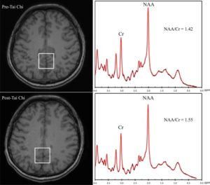
In today’s Research News article “Tai Chi Improves Brain Metabolism and Muscle Energetics in Older Adults.” (See summary below or view the full text of the study at: https://www.ncbi.nlm.nih.gov/pmc/articles/PMC6055800/ ), Zhou and colleagues recruited healthy older people (>55 years of age) and trained them in Tai Chi practice with a one hour, twice a week, for 12 weeks instruction. They were measured before and after training for leg strength while having their brains scanned for metabolites with Magnetic Resonance Imaging (MRI). N‐acetylaspartate (NAA) is a neuronal marker of neuronal health. They also measured the rate of recovery of phosphocreatine (PCr) in the leg following exercise, a marker of muscular fitness.


They found that after training there was a significant increase in N‐acetylaspartate (NAA). NAA is a marker of the number of neurons present in the brain. Hence it’s increase in this study suggests that Tai Chi training increases the number of brain cells in the elderly. This further suggests that Tai Chi training is neuroprotective and may reduce the degeneration of the brain that occurs in normal aging.


They also found that after training there was a significant decrease in the rate of recovery of phosphocreatine (PCr) in the leg following exercise. A PCr decrease indicates that the capacity of muscles to use oxygen has increased. This, then, is a measure of muscular fitness. Hence it’s decrease in this study suggests that Tai Chi training improves exercise fitness in older adults helping to counter the age related decline in strength.


These results suggest the biochemical mechanisms that may underlie the ability of Tai Chi training to slow or delay physical and mental decline and to increase brain matter. These results not only further support the benefits of Tai Chi training for aging adults but also indicate how this training may change the chemistry of the brain and muscles to counter the effects of aging.


So, improve brain metabolism and muscle energetics in older adults with Tai Chi.


CMCS – Center for Mindfulness and Contemplative Studies


This and other Contemplative Studies posts are also available on Google+ https://plus.google.com/106784388191201299496/posts and on Twitter @MindfulResearch


“Studies have shown that the incorporation of Tai Chi to an elders’ exercise program can be beneficial. Tai Chi practice was “beneficial to improve the balance control ability and flexibility of older adults, which may be the reason of preventing falls.” – Eric Edelman


In today’s Research News article “Tai Chi Improves Brain Metabolism and Muscle Energetics in

Study Summary


Zhou, M., Liao, H., Sreepada, L. P., Ladner, J. R., Balschi, J. A., & Lin, A. P. (2018). Tai Chi Improves Brain Metabolism and Muscle Energetics in Older Adults. Journal of Neuroimaging, 28(4), 359–364. http://doi.org/10.1111/jon.12515


ABSTRACT

BACKGROUND AND PURPOSE

Tai Chi is a mind‐body exercise that has been shown to improve both mental and physical health. As a result, recent literature suggests the use of Tai Chi to treat both physical and psychological disorders. However, the underlying physiological changes have not been characterized. The aim of this pilot study is to assess the changes in brain metabolites and muscle energetics after Tai Chi training in an aging population using a combined brain‐muscle magnetic resonance spectroscopy (MRS) examination.

METHODS

Six healthy older adults were prospectively recruited and enrolled into a 12‐week Tai Chi program. A brain 1H MRS and a muscle 31P MRS were scanned before and after the training, and postprocessed to measure N‐acetylaspartate to creatine (NAA/Cr) ratios and phosphocreatine (PCr) recovery time. Wilcoxon‐signed rank tests were utilized to assess the differences between pre‐ and post‐Tai Chi training.

RESULTS

A significant within‐subject increase in both the NAA/Cr ratios (P = .046) and the PCr recovery time (P =.046) was observed between the baseline and the posttraining scans. The median percentage changes were 5.38% and 16.51% for NAA/Cr and PCr recovery time, respectively.

CONCLUSIONS

Our pilot study demonstrates significant increase of NAA/Cr ratios in posterior cingulate gyrus and significantly improved PCr recovery time in leg muscles in older adults following short‐term Tai Chi training, and thus provides insight into the beneficial mechanisms.

https://www.ncbi.nlm.nih.gov/pmc/articles/PMC6055800/


Slow Mental Decline in the Elderly with Tai Chi

Slow Mental Decline in the Elderly with Tai Chi


By John M. de Castro, Ph.D.


“improvement of heart function combined with increased muscular power meant that the martial art should be considered the preferred technique for elderly people to maintain good health.” – The Telegraph


We celebrate the increasing longevity of the population. But, aging is a mixed blessing. The aging process involves a systematic progressive decline of the body and the brain. Every system in the body deteriorates including cognitive function (thinking ability) and motor function with a decline in strength, flexibility, and balance. It is inevitable. In addition, many elderly experience withdrawal and isolation from social interactions. There is some hope as there is evidence that these declines can be slowed. For example, a healthy diet and a regular program of exercise can slow the physical and cognitive decline of the body with aging. Also, contemplative practices such as meditation, yoga, and tai chi or qigong have all been shown to be beneficial in slowing or delaying physical and mental decline.


Tai Chi has been practiced for thousands of years with benefits for health and longevity. Tai Chi training is designed to enhance function and regulate the activities of the body through regulated breathing, mindful concentration, and gentle movements. Only recently though have the effects of Tai Chi practice been scrutinized with empirical research and found to be effective for an array of physical and psychological issues. Tai Chi has been shown to help the elderly improve attentionbalance, reducing fallsarthritiscognitive functionmemory, and reduce age related deterioration of the brain. Because Tai Chi is not strenuous, involving slow gentle movements, and is safe, having no appreciable side effects, it is appropriate for all ages including the elderly and for individuals with illnesses that limit their activities or range of motion.


In today’s Research News article “Tai Chi Improves Cognition and Plasma BDNF in Older Adults with Mild Cognitive Impairment: A Randomized Controlled Trial.”  Sungkarat and colleagues recruited elderly participants with mild cognitive impairments and randomly assigned them to either receive educational instruction related to cognitive impairment and fall prevention or practice Tai Chi at home guided by a 50-minute video, 3 times per week, for 6 months. They were measured at the beginning and end of training for cognitive performance, including memory, visuospatial ability, and executive function, and plasma markers for inflammation and neuroprotection, including plasma BDNF, TNF-α, and IL-10 levels.


They found that compared to baseline and control participants, the elderly who practiced Tai Chi had significantly improved levels of cognitive function, including improvements in memory and thinking ability (executive function). In addition, Tai Chi practice was found to significantly increase the levels of brain-derived neurotrophic factor (BDNF). BDNF is a neurotrophic factor that works to protect the brain from deterioration and promote the growth of brain cells. Hence, they found that Tai Chi practice reduces cognitive decline with aging and increases neuroprotection.


The cognitive decline with aging has been associated with degeneration of neural tissues. On the other hand, mindfulness practices have been found to change the brain and protect it against age related decline. The present results add further evidence that mindfulness practices, Tai Chi  in particular, improves memory and cognitive performance and promotes neuroprotection in the elderly. The attractiveness of the low intensity, low cost, convenient, and socially fun nature of Tai Chi practice makes it a great treatment for the prevention of age related decline.


So, slow mental decline in the elderly with Tai Chi.


“Scientists . . . found increases in brain volume and improvements on tests of memory and thinking in Chinese seniors who practiced Tai Chi three times a week.” – Science Daily


CMCS – Center for Mindfulness and Contemplative Studies


This and other Contemplative Studies posts are also available on Google+ https://plus.google.com/106784388191201299496/posts and on Twitter @MindfulResearch


Study Summary


Somporn Sungkarat, PhD, SirinunBoripuntakul, PhD, Sirinart Kumfu, PhD, Stephen R. Lord, PhD, Nipon Chattipakorn, MD, PhD. Tai Chi Improves Cognition and Plasma BDNF in Older Adults With Mild Cognitive Impairment: A Randomized Controlled Trial. Neurorehabilitation and Neural Repair. First Published January 20, 2018, https://doi-org.ezproxy.shsu.edu/10.1177/15459683177536


Abstract

Background. Effects of Tai Chi (TC) on specific cognitive function and mechanisms by which TC may improve cognition in older adults with amnestic mild cognitive impairment (a-MCI) remain unknown. Objective. To examine the effects of TC on cognitive functions and plasma biomarkers (brain-derived neurotrophic factor [BDNF], tumor necrosis factor-α [TNF-α], and interleukin-10 [IL-10]) in a-MCI. Methods. A total of 66 older adults with a-MCI (mean age = 67.9 years) were randomized to either a TC (n = 33) or a control group (n = 33). Participants in the TC group learned TC with a certified instructor and then practiced at home for 50 min/session, 3 times/wk for 6 months. The control group received educational material that covered information related to cognition. The primary outcome was cognitive performance, including Logical Memory (LM) delayed recall, Block Design, Digit Span, and Trail Making Test B minus A (TMT B-A). The secondary outcomes were plasma biomarkers, including BDNF, TNF-α, and IL-10. Results. At the end of the trial, performance on the LM and TMT B-A was significantly better in the TC group compared with the control group after adjusting for age, gender, and education (P < .05). Plasma BDNF level was significantly increased for the TC group, whereas the other outcome measures were similar between the 2 groups after adjusting for age and gender (P < .05). Conclusions. TC training significantly improved memory and the mental switching component of executive function in older adults with a-MCI, possibly via an upregulation of BDNF.

http://journals.sagepub.com.ezproxy.shsu.edu/doi/full/10.1177/1545968317753682

Change Major Depression Brain Chemistry with Mindfulness

MBCT Major Depression2 Li

By John M. de Castro, Ph.D.


“Mindfulness-based cognitive therapy helps participants in the classes to see more clearly the patterns of the mind; and to learn how to recognize when their mood is beginning to go down. It helps break the link between negative mood and the negative thinking that it would normally have triggered. Participants develop the capacity to allow distressing mood, thoughts and sensations to come and go, without having to battle with them. They find that they can stay in touch with the present moment, without having to ruminate about the past, or worry about the future.” – Center for Suicide Research


Major Depressive Disorder (MDD) is a severe mood disorder that includes mood dysregulation and cognitive impairment. It is estimated that 16 million adults in the U.S. (6.9% of the population suffered from major depression in the past year and affects females (8.4%) to a great extent than males (5.2%). It’s the second-leading cause of disability in the world following heart disease. It has also been shown that depression is, to a large extent, inherited, but can also be affected by the environment. Since the genes basically encode when, where, and how chemicals are produced, it is likely that there are changes in brain chemistry produced by the genes responsible for Major Depressive Disorder.


The usual treatment of choice for MDD is drug treatment. This supports the altered brain chemistry notion for MDD since the most effective treatment for MDD, drug treatment, changes brain chemistry. In fact, it is estimated that 10% of the U.S. population is taking some form of antidepressant medication. But a substantial proportion of patients (~40%) do not respond to drug treatment. In addition, the drugs can have nasty side effects. So, there is need to explore other treatment options.


Mindfulness meditation is a safe alternative that has been shown to be effective for major depressive disorder even in individuals who do not respond to drug treatment. Mindfulness Based Cognitive Therapy (MBCT) was developed specifically to treat depression and has been shown to be very effective in treating existing depression and preventing relapse when depression is in remission. It makes sense that if altered brain chemistry underlies MDD and that MBCT is an effective treatment for MDD, then MBCT must in some way change brain chemistry. In today’s Research News article “Evaluating metabolites in patients with major depressive disorder who received mindfulness-based cognitive therapy and healthy controls using short echo MRSI at 7 Tesla”

https://www.facebook.com/ContemplativeStudiesCenter/photos/a.628903887133541.1073741828.627681673922429/1196100317080559/?type=3&theater

Li and colleagues explore brain chemistry changes in Major Depressive Disorder (MDD) and the effects of Mindfulness Based Cognitive Therapy (MBCT) on those brain chemistry changes.


They recruited patients who were diagnosed with MDD but who were not currently taking antidepressant drugs and who were not practicing meditation or yoga. The brains of these patients and healthy controls were scanned with a powerful imaging technique called Magnetic Resonance Spectroscopic Imaging (MRSI). It is capable of non-invasively detecting levels of particular chemicals in the brain. The patients then received an 8-week MBCT group therapy followed by rescanning of the brains for the same chemicals.


They found that the MDD patients compared to healthy controls had elevated levels of choline-containing compounds and decreased levels of N-acetyl aspartate, myo-inositol, and glutathione.

These chemicals are breakdown products of active brain chemicals (metabolites). These are all markers of brain function. The heightened levels of choline-containing compounds suggests that there is with increased cell density and/or membrane turnover in MDD. The decreased levels of N-acetyl aspartate suggest that there is a loss of neurons or neuronal function in MDD. The decreased levels of myo-inositol suggest that there is a loss of or dysfunction of glial cells in MDD. Finally, the decreased levels of glutathione suggest that there is a lower level of neuron excitation in the brain in MDD.


Importantly, Li and colleagues found that MBCT significantly reduced depression levels and at the same time normalized the levels of all of the metabolites that had abnormal levels in the patients. These are potentially important results. They demonstrate altered brain chemistry in MDD suggestive of dysfunction in the normal activities of the nervous system and point to potential causal factors in MDD. They also provide suggestions as to how MBCT changes the brain to effectively treat MDD.


It should be noted that the changes in metabolites in Major Depressive Disorder may be the result of the depression rather than its cause. The fact that the changes vanished after treatment reduced depression tends to support this contention. It is a complex disease effecting the most complex entity in the universe, the human brain. Hence, there is still a lot of work to do to determine the causal factors in MDD.


Regardless, change major depression brain chemistry with mindfulness.


 “Mindfulness is the only thing I know to do that can dig me out of despair and give me even a few seconds of time out from me,” – Ruby Wax


CMCS – Center for Mindfulness and Contemplative Studies