Less Complex Brain Activity Characterizes Meditation by Experienced Meditators.

Less Complex Brain Activity Characterizes Meditation by Experienced Meditators.


By John M. de Castro, Ph.D.


Using modern technology like fMRI scans, scientists have developed a more thorough understanding of what’s taking place in our brains when we meditate. The overall difference is that our brains stop processing information as actively as they normally would.” – Belle Beth Cooper


There has accumulated a large amount of research demonstrating that meditation practice has significant benefits for psychological, physical, and spiritual wellbeing. One way that meditation practices may produce these benefits is by altering the brain. The nervous system is a dynamic entity, constantly changing and adapting to the environment. It will change size, activity, and connectivity in response to experience. These changes in the brain are called neuroplasticity. Over the last decade neuroscience has been studying the effects of contemplative practices on the brain and has identified neuroplastic changes in widespread areas. In other words, meditation practice appears to mold and change the brain, producing psychological, physical, and spiritual benefits.


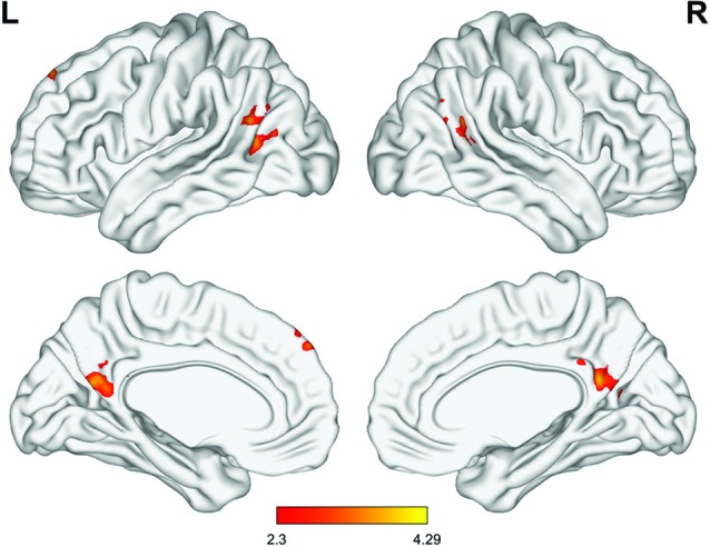
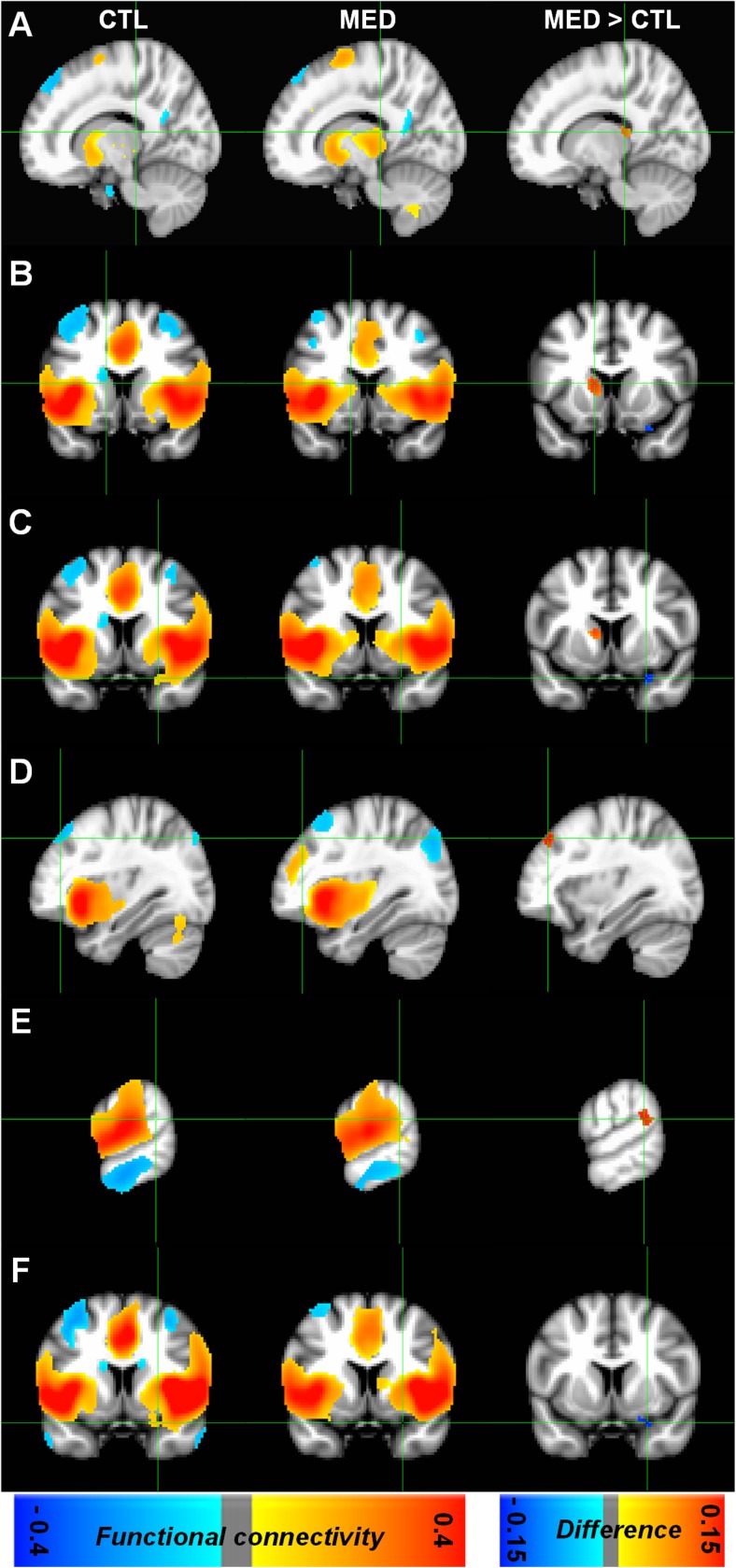
It is important to understand what are the exact changes in the brain that are produced by meditation. In today’s Research News article “Characterizing the Dynamical Complexity Underlying Meditation.” (See summary below or view the full text of the study at: https://www.ncbi.nlm.nih.gov/pmc/articles/PMC6637306/), Escrichs and colleagues recruited experienced adult meditators with at least 1000 hours of meditation experience and an ongoing practice and a matched group of non-meditators. They underwent functional Magnetic Resonance Imaging (fMRI) at rest and again when performing breath focused meditation. The scans were then analyzed with Intrinsic Ignition Framework that measures the degree of elicited whole-brain integration of spontaneously occurring events across time, in other words the complexity of information processing going on in the nervous system.


They found that at rest, the meditators had higher Intrinsic-Driven Mean Integration (IDMI) than controls but during meditation they had significantly lower IDMI than the controls. The meditators also had significantly higher metastability during rest than controls but that metastability significantly declined during meditation. These results are complex but indicate that meditators have greater levels of information moving around the brain and greater complexity of information processing over time at rest but during meditation move to a state where there is less information moving around and less complexity of processing.


The results suggest that meditators have more complicated information processing going on in their nervous systems at rest but during meditation greatly simplify that activity. It would appear that this takes practice as the non-meditators did not have comparable activities during meditation. This suggests that meditation experience over time produces neuroplastic alterations of the brain that increase the ability of the brain to process information normally and to become quieter during meditation.


Nondirective meditation yields more marked changes in electrical brain wave activity associated with wakeful, relaxed attention, than just resting without any specific mental technique.” – ScienceDaily

CMCS – Center for Mindfulness and Contemplative Studies


This and other Contemplative Studies posts are also available on Google+ https://plus.google.com/106784388191201299496/posts and on Twitter @MindfulResearch


Study Summary


Escrichs, A., Sanjuán, A., Atasoy, S., López-González, A., Garrido, C., Càmara, E., & Deco, G. (2019). Characterizing the Dynamical Complexity Underlying Meditation. Frontiers in systems neuroscience, 13, 27. doi:10.3389/fnsys.2019.00027


Abstract

Over the past 2,500 years, contemplative traditions have explored the nature of the mind using meditation. More recently, neuroimaging research on meditation has revealed differences in brain function and structure in meditators. Nevertheless, the underlying neural mechanisms are still unclear. In order to understand how meditation shapes global activity through the brain, we investigated the spatiotemporal dynamics across the whole-brain functional network using the Intrinsic Ignition Framework. Recent neuroimaging studies have demonstrated that different states of consciousness differ in their underlying dynamical complexity, i.e., how the broadness of communication is elicited and distributed through the brain over time and space. In this work, controls and experienced meditators were scanned using functional magnetic resonance imaging (fMRI) during resting-state and meditation (focused attention on breathing). Our results evidenced that the dynamical complexity underlying meditation shows less complexity than during resting-state in the meditator group but not in the control group. Furthermore, we report that during resting-state, the brain activity of experienced meditators showed higher metastability (i.e., a wider dynamical regime over time) than the one observed in the control group. Overall, these results indicate that the meditation state operates in a different dynamical regime compared to the resting-state.

https://www.ncbi.nlm.nih.gov/pmc/articles/PMC6637306/


Structural and Functional Changes in the Brain Produced by Meditation Training

Structural and Functional Changes in the Brain Produced by Meditation Training


By John M. de Castro, Ph.D.


“Measurable changes in brain regions associated with memory, sense of self, empathy, and stress start to appear in subjects who practice mindfulness meditation for only eight weeks.” – Deepak Chopra


There has accumulated a large amount of research demonstrating that mindfulness has significant benefits for psychological, physical, and spiritual wellbeing. One way that mindfulness practices may produce these benefits is by altering the brain. The nervous system is a dynamic entity, constantly changing and adapting to the environment. It will change size, activity, and connectivity in response to experience. These changes in the brain are called neuroplasticity. Over the last decade neuroscience has been studying the effects of contemplative practices on the brain and has identified neuroplastic changes in widespread areas. In other words, mindfulness practice appears to mold and change the brain, producing psychological, physical, and spiritual benefits.


Although, these changes have been documented, there is scant evidence regarding the temporal course of the neural changes with increasing experience with meditation. In today’s Research News article “Alterations in Brain Structure and Amplitude of Low-frequency after 8 weeks of Mindfulness Meditation Training in Meditation-Naïve Subjects.” (See summary below or view the full text of the study at: https://www.ncbi.nlm.nih.gov/pmc/articles/PMC6662752/), Yang and colleagues recruited meditation naïve college students and provided them with a 8 week meditation training program. They met for 1.5 hours once a week and were requested to meditate at home for 45 minutes daily. They were measured before and after the meditation program for mindfulness, anxiety, depression, and mood, including anger, fatigue, tension, depression, vigour and friendliness. In addition, their brains were measured with functional Magnetic Resonance Imaging (fMRI) before and after training.


They found that after meditation training there were significant decreases in anxiety and depression and significant increases in the non-reactivity facet of mindfulness. Cortical thickness significantly increased over training in the precuneus and superior parietal lobule while local brain activity fluctuations decreased in the precuneus and inferior parietal lobule. The parietal cortex is associated with bodily sensation and self-referential thinking while the precuneus is associated with the default mode network involved in mind wandering and self-referential thought.


The study did not contain a control condition. So, conclusion must be reached carefully. But the results suggest that mindfulness meditation training decreases anxiety and depression, which has been previously well documented. The neural findings that meditation training resulted in decreased brain activity fluctuations in the precuneus and inferior parietal lobule suggests that the training reduces activity in brain regions associated with mind wandering and self-referential thinking, which have also been well documented previously. Focusing on the present moment as is trained in mindfulness meditation cannot coexist with mind wandering and self-referential thinking. So, it is not surprising that there’s reduced activity in the brain regions underlying these functions.


All of this suggests that mindfulness meditation training changes the brain in ways that reflect greater present moment awareness and less daydreaming and discursive thinking.


“as the popularity of mindfulness grows, brain imaging techniques are revealing that this ancient practice can profoundly change the way different regions of the brain communicate with each other – and therefore how we think – permanently.” – Tom Ireland


CMCS – Center for Mindfulness and Contemplative Studies


This and other Contemplative Studies posts are also available on Google+ https://plus.google.com/106784388191201299496/posts and on Twitter @MindfulResearch


Study Summary


Chuan-Chih Yang, Alfonso Barrós-Loscertales, Meng Li, Daniel Pinazo, Viola Borchardt, César Ávila, Martin Walter. Alterations in Brain Structure and Amplitude of Low-frequency after 8 weeks of Mindfulness Meditation Training in Meditation-Naïve Subjects. Sci Rep. 2019; 9: 10977. Published online 2019 Jul 29. doi: 10.1038/s41598-019-47470-4


Abstract

Increasing neuroimaging evidence suggests that mindfulness meditation expertise is related to different functional and structural configurations of the default mode network (DMN), the salience network (SN) and the executive network at rest. However, longitudinal studies observing resting network plasticity effects in brains of novices who started to practice meditation are scarce and generally related to one dimension, such as structural or functional effects. The purpose of this study was to investigate structural and functional brain network changes (e.g. DMN) after 40 days of mindfulness meditation training in novices and set these in the context of potentially altered depression symptomatology and anxiety. We found overlapping structural and functional effects in precuneus, a posterior DMN region, where cortical thickness increased and low-frequency amplitudes (ALFF) decreased, while decreased ALFF in left precuneus/posterior cingulate cortex correlates with the reduction of (CES-D) depression scores. In conclusion, regional overlapping of structural and functional changes in precuneus may capture different components of the complex changes of mindfulness meditation training.

https://www.ncbi.nlm.nih.gov/pmc/articles/PMC6662752/


Mindfulness is Associated with Improved Functional Connectivity in the Brain

Mindfulness is Associated with Improved Functional Connectivity in the Brain


By John M. de Castro, Ph.D.


“mindfulness meditation training increases resting state connectivity between top-down executive control regions, highlighting an important mechanism through which it reduces stress levels.” – Daniel Reed


The nervous system is a dynamic entity, constantly changing and adapting to the environment. It will change size, activity, and connectivity in response to experience. These changes in the brain are called neuroplasticity.  Over the last decade neuroscience has been studying the effects of contemplative practices on the brain and has identified neuroplastic changes in widespread area. and have found that meditation practice appears to mold and change the brain, producing psychological, physical, and spiritual benefits.


This suggests that the individual’s trait of mindfulness may be associated with differing functional connectivity in the brain. In today’s Research News article “Trait Mindfulness and Functional Connectivity in Cognitive and Attentional Resting State Networks.” (See summary below or view the full text of the study at: https://www.ncbi.nlm.nih.gov/pmc/articles/PMC6473082/), Parkinson and colleagues recruited undergraduate students and measured them for mindfulness including the observing, describing, non-reacting, acting with awareness, and non-judging facets. Functional connectivity of their brains was measured with Magnetic Resonance Imaging (MRI).


They found that the higher the students’ levels of trait mindfulness and its facets the lower the functional connectivity in a set of structures termed the Default Mode Network (DMN) that has been associated with mind wandering, rumination, and self-referential thinking. Conversely, they found that the higher the students’ levels of trait mindfulness the higher the functional connectivity of the Anterior Cingulate Cortex, a structure associated with self-regulation, the Dorsal Medial Prefrontal Cortex, a structure associated with attentional control and high level thinking (executive function), the Insula, a structure associated with emotion regulation, and the Prefrontal Gyrus, a structure associated with sensorimotor processing.


Hence they found that there was increased functional connectivity in structures that appear to underlie the relationships of mindfulness with attention, emotion regulation, sensory processing, self-regulation, and high level thinking and decreased functional connectivity in structures that appear to underlie the processes of mind wandering and rumination that are weakened with mindfulness. In many ways, the functional connectivity of the students with high mindfulness resembles that of experienced meditators. This suggests that mindfulness and its benefits are associated with strengthened neural processing in specific areas of the brain.


Hence, mindfulness is associated with improved functional connectivity in the brain.


Just 11 hours of learning a meditation technique induce positive structural changes in brain connectivity by boosting efficiency in a part of the brain that helps a person regulate behavior in accordance with their goals.” – University of Oregon


CMCS – Center for Mindfulness and Contemplative Studies


This and other Contemplative Studies posts are also available on Google+ https://plus.google.com/106784388191201299496/posts and on Twitter @MindfulResearch


Study Summary


Parkinson TD, Kornelsen J, Smith SD. Trait Mindfulness and Functional Connectivity in Cognitive and Attentional Resting State Networks. Front Hum Neurosci. 2019 Apr 12;13:112. doi: 10.3389/fnhum.2019.00112. PubMed PMID: 31031607; PubMed Central PMCID: PMC6473082.


Abstract

Mindfulness has been described as an orienting of attention to the present moment, with openness and compassion. Individuals displaying high trait mindfulness exhibit this tendency as a more permanent personality attribute. Given the numerous physical and mental health benefits associated with mindfulness, there is a great interest in understanding the neural substrates of this trait. The purpose of the current research was to examine how individual differences in trait mindfulness associated with functional connectivity in five resting-state networks related to cognition and attention: the default mode network (DMN), the salience network (SN), the central executive network (CEN), and the dorsal and ventral attention networks (DAN and VAN). Twenty-eight undergraduate participants completed the Five-Facet Mindfulness Questionnaire (FFMQ), a self-report measure of trait mindfulness which also provides scores on five of its sub-categories (Observing, Describing, Acting with Awareness, Non-judging of Inner Experience, and Non-reactivity to Inner Experience). Participants then underwent a structural MRI scan and a 7-min resting state functional MRI scan. Resting-state data were analyzed using independent-component analyses. An analysis of covariance (ANCOVA) was performed to determine the relationship between each resting state network and each FFMQ score. These analyses indicated that: (1) trait mindfulness and its facets showed increased functional connectivity with neural regions related to attentional control, interoception, and executive function; and (2) trait mindfulness and its facets showed decreased functional connectivity with neural regions related to self-referential processing and mind wandering. These patterns of functional connectivity are consistent with some of the benefits of mindfulness—enhanced attention, self-regulation, and focus on present experience. This study provides support for the notion that non-judgmental attention to the present moment facilitates the integration of regions in neural networks that are related to cognition, attention, and sensation.

https://www.ncbi.nlm.nih.gov/pmc/articles/PMC6473082/


Improve Caregiver Psychological Health by Changing the Brain Response with Mindfulness

Improve Caregiver Psychological Health by Changing the Brain Response with Mindfulness


By John M. de Castro, Ph.D.


“We are set up for short-term stress, but caregiving is long-term stress. Mindfulness is basically coming back into the present moment, so it works to inhibit the stress response. Most of us run around listening to our thoughts, and this is particularly true of caregivers, who are driven by the to-do list. They are never at rest.” – Joan Griffiths Vega


Dementia is a progressive loss of mental function produced by degenerative diseases of the brain. Dementia patients require caregiving particularly in the later stages of the disease. Caregiving for dementia patients is a daunting intense experience that can go on for four to eight years with increasing responsibilities as the loved one deteriorates. This places tremendous psychological and financial stress on the caregiver. Hence, there is a need to both care for the dementia patients and also for the caregivers. Mindfulness practice for caregivers has been shown to help them cope with the physical and psychological demands of caregiving. In addition, mindfulness training has been found to help protect aging individuals from physical and cognitive declines.


The nervous system is a dynamic entity, constantly changing and adapting to the environment. It will change size, activity, and connectivity in response to experience. These changes in the brain are called neuroplasticity.  Over the last decade neuroscience has been studying the effects of contemplative practices on the brain and has identified neuroplastic changes in widespread area. and have found that meditation practice appears to mold and change the brain, producing psychological, physical, and spiritual benefits.


In today’s Research News article “Grief, Mindfulness and Neural Predictors of Improvement in Family Dementia Caregivers.” (See summary below or view the full text of the study at: https://www.ncbi.nlm.nih.gov/pmc/articles/PMC6530345/), Jain and colleagues recruited dementia caregivers (91% female) and provided for them either 4 weeks of mindfulness training or 4 weeks of relaxation training. Training occurred in a once a week meeting along with home practice. The participants were measured before and after training for grief, depression, and mindfulness. They then had brain scans performed with functional Magnetic Resonance Imaging (fMRI) while they viewed pictures of their loved one with dementia or a stranger with the pictures labelled with either grief related words, e.g. disease, dementia, and sick or with neutral words e.g. village, planter and curve.


They found that at baseline grief and depression levels were high and strongly related. They also found that the higher the levels of mindfulness the lower the levels of grief and depression. After mindfulness training there were reductions in grief and depression and increases in mindfulness. These findings are similar to previous research of improvements in the mental health of caregivers after mindfulness training.


Interestingly, in comparison to pictures of strangers, when showed pictures of their loved ones with dementia the caregivers showed increases in brain activation in the dorsal anterior cingulate gyrus and precuneus. Viewing grief related words results in increased activity in the medial prefrontal cortex. The greater the decreases in grief following training the greater the activation of the medial prefrontal cortex and precuneus.


The structures showing activation to the caregiver’s loved one all are components of what is called the default mode network which is involved in self-referential thinking and thinking about others. It would appear that the mindfulness training resulted in greater thinking about the dementia patient and the self when viewing a picture of the patient. This may be reflective of heightened compassion for the self and the patient. This in turn, may produce improvements in the caregivers mental health.


So, improve caregiver psychological health by changing the brain response with mindfulness.


CMCS – Center for Mindfulness and Contemplative Studies


This and other Contemplative Studies posts are also available on Google+ https://plus.google.com/106784388191201299496/posts and on Twitter @MindfulResearch


“By focusing on the fact that families and communities are producers of health and health care, not just clients or consumers, it empowers families and communities to co-create health interventions,” – Alicia Bazzano


Study Summary


Jain, F. A., Connolly, C. G., Moore, L. C., Leuchter, A. F., Abrams, M., Ben-Yelles, R. W., … Iacoboni, M. (2019). Grief, Mindfulness and Neural Predictors of Improvement in Family Dementia Caregivers. Frontiers in human neuroscience, 13, 155. doi:10.3389/fnhum.2019.00155


Abstract

Background: Family dementia caregivers often suffer from an immense toll of grief while caring for their loved ones. We sought to identify the clinical relationship between grief, depression and mindfulness and identify neural predictors of symptomatology and improvement.

Methods: Twenty three family dementia caregivers were assessed at baseline for grief, mindfulness and depression, of which 17 underwent functional magnetic resonance imaging (fMRI). During fMRI, caregivers were shown faces of either their dementia-stricken relative or that of a stranger, paired with grief-related or neutral words. In nine subjects, post fMRI scans were also obtained after 4 weeks of either guided imagery or relaxation. Robust regression was used to predict changes in symptoms with longitudinal brain activation (BA) changes as the dependent variable.

Results: Grief and depression symptoms were correlated (r = 0.50, p = 0.01), and both were negatively correlated with mindfulness (r = −0.70, p = 0.0002; r = −0.52, p = 0.01). Relative to viewing strangers, caregivers showed pictures of their loved ones (picture factor) exhibited increased activation in the dorsal anterior cingulate gyrus and precuneus. Improvement in grief but not mindfulness or depression was predicted by increased relative BA in the precuneus and anterior cingulate (different subregions from baseline). Viewing grief-related vs. neutral words elicited activity in the medial prefrontal cortex and precuneus.

Conclusions: Caregiver grief, depression and mindfulness are interrelated but have at least partially nonoverlapping neural mechanisms. Picture and word stimuli related to caregiver grief evoked brain activity in regions previously identified with bereavement grief. These activation foci might be useful as biomarkers of treatment response.

https://www.ncbi.nlm.nih.gov/pmc/articles/PMC6530345/

Alter the Brain to Deal with Stress with Meditation and Yoga

Alter the Brain to Deal with Stress with Meditation and Yoga


By John M. de Castro, Ph.D.


“Brain researchers have detected improvements in cognition and emotional well-being associated with meditation and yoga, as well as differences in how meditation and prayer affect the brain.” – Michaela Jarvis


There has accumulated a large amount of research demonstrating that mindfulness has significant benefits for psychological, physical, and spiritual wellbeing. One way that mindfulness practices may produce these benefits is by altering the brain. The nervous system is a dynamic entity, constantly changing and adapting to the environment. It will change size, activity, and connectivity in response to experience. These changes in the brain are called neuroplasticity. Over the last decade neuroscience has been studying the effects of contemplative practices on the brain and has identified neuroplastic changes in widespread areas. In other words, mindfulness practice appears to mold and change the brain, producing psychological, physical, and spiritual benefits.


In today’s Research News article “Meditation and yoga practice are associated with smaller right amygdala volume: the Rotterdam study.” (See summary below or view the full text of the study at: https://www.ncbi.nlm.nih.gov/pmc/articles/PMC6302143/), Gotink and colleagues utilized the data on participants in the longitudinal Rotterdam Study who were 45 years of age and older  at the time of recruitment and at the time of measurement had a mean age of 64 years. They were interviewed to determine if the practiced meditation and yoga and whether these practices improved their coping with stress. They were also measured for body size, blood pressure, blood fat, diabetes, smoking, alcohol use, stress, anxiety, and depression. In addition, their brains were scanned with Magnetic Resonance Imaging (MRI).


They found that practitioners of meditation and yoga reported higher stress levels than non-practitioners, but reported that the practice helped them cope with the stress. In addition, meditation practitioners had higher depression levels than non-practitioners. It is possible that people who are under high levels of stress or are depressed tend to engage in meditation and yoga practices to help cope with it.


They also report that the practitioners had smaller volumes of the brain structures right side amygdala and left hippocampus. In addition, over a five-year period the practitioners had a significant decrease in amygdala volume. The amygdala is associated with negative emotions and its smaller volume may suggest fewer or weaker negative emotions in practitioners.


This was a cross-sectional study and causation cannot be determined. It is possible that people with certain types of brains are more likely to practice. It will require a randomized controlled trial to determine what effects yoga and meditation practice may have on the psychological state and nervous system volumes.


Alter the brain to deal with stress with meditation and yoga.


“Studies show that yoga increases relaxation in the brain, improves areas of the brain that help us manage pain, and protects us against age-related decline. Together, these benefits begin to reveal the scientifically validated effects of yoga practice on brain health.” – Angela Wilson


CMCS – Center for Mindfulness and Contemplative Studies


This and other Contemplative Studies posts are also available on Google+ https://plus.google.com/106784388191201299496/posts and on Twitter @MindfulResearch


Study Summary


Gotink, R. A., Vernooij, M. W., Ikram, M. A., Niessen, W. J., Krestin, G. P., Hofman, A., … Hunink, M. (2018). Meditation and yoga practice are associated with smaller right amygdala volume: the Rotterdam study. Brain imaging and behavior, 12(6), 1631–1639. doi:10.1007/s11682-018-9826-z


Abstract

To determine the association between meditation and yoga practice, experienced stress, and amygdala and hippocampal volume in a large population-based study. This study was embedded within the population-based Rotterdam Study and included 3742 participants for cross-sectional association. Participants filled out a questionnaire assessing meditation practice, yoga practice, and experienced stress, and underwent a magnetic resonance scan of the brain. 2397 participants underwent multiple brain scans, and were assessed for structural change over time. Amygdala and hippocampal volumes were regions of interest, as these are structures that may be affected by meditation. Multivariable linear regression analysis and mixed linear models were performed adjusted for age, sex, educational level, intracranial volume, cardiovascular risk, anxiety, depression and stress. 15.7% of individuals participated in at least one form of practice. Those who performed meditation and yoga practices reported significantly more stress (mean difference 0.2 on a 1–5 scale, p < .001) and more depressive symptoms (mean difference 1.03 on CESD, p = .015). Partaking in meditation and yoga practices was associated with a significantly lower right amygdala volume (β = − 31.8 mm3, p = .005), and lower left hippocampus volume (β = − 75.3 mm3, p = .025). Repeated measurements using linear mixed models showed a significant effect over time on the right amygdala of practicing meditation and yoga (β = − 24.4 mm3, SE 11.3, p = .031). Partaking in meditation and yoga practice is associated with more experienced stress while it also helps cope with stress, and is associated with smaller right amygdala volume.

https://www.ncbi.nlm.nih.gov/pmc/articles/PMC6302143/


Change Brain Connectivity for Better Attention and Thinking with Mindfulness

Change Brain Connectivity for Better Attention and Thinking with Mindfulness


By John M. de Castro, Ph.D.


“mindfulness meditation training increases resting state connectivity between top-down executive control regions, highlighting an important mechanism through which it reduces stress levels.” – Daniel Reed


There has accumulated a large amount of research demonstrating that mindfulness has significant benefits for psychological, physical, and spiritual wellbeing. It even improves high level thinking known as executive function and emotion regulation and compassion. Its positive effects are so widespread that it is difficult to find any other treatment of any kind with such broad beneficial effects on everything from thinking to mood and happiness to severe mental and physical illnesses. This raises the question of how mindfulness training could produce such widespread and varied benefits. One possibility is that mindfulness practice results in beneficial changes in the nervous system.


The nervous system is a dynamic entity, constantly changing and adapting to the environment. It will change size, activity, and connectivity in response to experience. These changes in the brain are called neuroplasticity. Over the last decade neuroscience has been studying the effects of contemplative practices on the brain and has identified neuroplastic changes in widespread areas. In other words, mindfulness practice appears to mold and change the brain, producing psychological, physical, and spiritual benefits. The changes are complex and require sophisticated brain scanning techniques to detect. Hence there is a need to continue investigating the nature of these changes in the brain produced by meditation.


In today’s Research News article “Trait Mindfulness and Functional Connectivity in Cognitive and Attentional Resting State Networks.” (See summary below or view the full text of the study at: https://www.ncbi.nlm.nih.gov/pmc/articles/PMC6473082/), Parkinson and colleagues recruited undergraduate students who had never meditated, measured them for mindfulness, and scanned their brains under resting conditions with functional Magnetic Resonance Imaging (fMRI). They examined the functional connectivity of a number of established neural networks and their relationship with mindfulness.


They found that mindfulness was negatively related to the functional connectivity of the Default Mode Network (DMN) and positively related to the functional connectivity of the Salience Network, the Central Executive Network, and Attention Network. The Default Mode Network (DMN) has been shown to be associated with mind wandering and self-referential thinking. It is not surprising that mindfulness would be associated with lower levels of the functioning of this network. Indeed, previous work has demonstrated that mindfulness is associated with reduced “mind wandering.”


The Salience Network is involved in detecting and filtering important stimuli in the environment from the environment and thereby gets involved in a myriad of high level psychological and social functions. The results suggest that being more mindful is associated with being more sensitive to important information.


The Central Executive Network has been shown to be associated with high level thinking and behavioral control. Hence, the results further suggest that high mindfulness is associated with improved cognition. Indeed, mindfulness has been shown through extensive research to be associated with better cognitive ability.


Finally, the Attention Network has been found to be associated with, no surprise, the ability to attend and focus. This suggests that high mindfulness is associated with improved attention ability. Again, this reflects other research which demonstrated that mindfulness is associated with a greater ability to attend.


Hence the study demonstrated the associations with mindfulness with functional connectivity in various neural networks tracks the demonstrated effects of mindfulness on the individual’s ability to focus, think, and stay in the present moment. This further suggests that changes in the operations of the brain are produced by mindfulness and that hese changes in turn produced improved functional capacities.


So, change brain connectivity for better attention and thinking with mindfulness.


“Just 11 hours of learning a meditation technique induce positive structural changes in brain connectivity by boosting efficiency in a part of the brain that helps a person regulate behavior in accordance with their goals,” – University of Oregon


CMCS – Center for Mindfulness and Contemplative Studies


This and other Contemplative Studies posts are also available on Google+ https://plus.google.com/106784388191201299496/posts and on Twitter @MindfulResearch


Study Summary


Parkinson, T. D., Kornelsen, J., & Smith, S. D. (2019). Trait Mindfulness and Functional Connectivity in Cognitive and Attentional Resting State Networks. Frontiers in Human Neuroscience, 13, 112. doi:10.3389/fnhum.2019.00112


Abstract

Mindfulness has been described as an orienting of attention to the present moment, with openness and compassion. Individuals displaying high trait mindfulness exhibit this tendency as a more permanent personality attribute. Given the numerous physical and mental health benefits associated with mindfulness, there is a great interest in understanding the neural substrates of this trait. The purpose of the current research was to examine how individual differences in trait mindfulness associated with functional connectivity in five resting-state networks related to cognition and attention: the default mode network (DMN), the salience network (SN), the central executive network (CEN), and the dorsal and ventral attention networks (DAN and VAN). Twenty-eight undergraduate participants completed the Five-Facet Mindfulness Questionnaire (FFMQ), a self-report measure of trait mindfulness which also provides scores on five of its sub-categories (Observing, Describing, Acting with Awareness, Non-judging of Inner Experience, and Non-reactivity to Inner Experience). Participants then underwent a structural MRI scan and a 7-min resting state functional MRI scan. Resting-state data were analyzed using independent-component analyses. An analysis of covariance (ANCOVA) was performed to determine the relationship between each resting state network and each FFMQ score. These analyses indicated that: (1) trait mindfulness and its facets showed increased functional connectivity with neural regions related to attentional control, interoception, and executive function; and (2) trait mindfulness and its facets showed decreased functional connectivity with neural regions related to self-referential processing and mind wandering. These patterns of functional connectivity are consistent with some of the benefits of mindfulness—enhanced attention, self-regulation, and focus on present experience. This study provides support for the notion that non-judgmental attention to the present moment facilitates the integration of regions in neural networks that are related to cognition, attention, and sensation.

https://www.ncbi.nlm.nih.gov/pmc/articles/PMC6473082/


Change the Brain to Improve Resilience with Meditation

Change the Brain to Improve Resilience with Meditation


By John M. de Castro, Ph.D.


“Mindful people … can better cope with difficult thoughts and emotions without becoming overwhelmed or shutting down (emotionally). Pausing and observing the mind may (help us) resist getting drawn into wallowing in a setback.” – Badri Bajaj


Psychological well-being is sometimes thought of as a lack of mental illness. But it is more than just a lack of something. It is a positive set of characteristics that lead to happy, well-adjusted life. These include the ability to be aware of and accept one’s strengths and weaknesses, to have goals that give meaning to life, to truly believe that your potential capabilities are going to be realized, to have close and valuable relations with others, the ability to effectively manage life issues especially daily issues, and the ability to follow personal principles even when opposed to society. But stress can interfere with the individual’s ability to achieve these goals.  When highly stressed, resilience is required to cope with the stress and continue on the path to psychological well-being.


One way that mindfulness practices such as meditation may improve resilience is by altering the brain. The nervous system is a dynamic entity, constantly changing and adapting to the environment. It will change size, activity, and connectivity in response to experience. These changes in the brain are called neuroplasticity. Over the last decade neuroscience has been studying the effects of contemplative practices on the brain and has identified neuroplastic changes in widespread areas. In other words, mindfulness practice appears to mold and change the brain, producing psychological, physical, and spiritual benefits. The changes in the brain, however, that are responsible for increased resilience are unknown.


In today’s Research News article “The Immediate and Sustained Positive Effects of Meditation on Resilience Are Mediated by Changes in the Resting Brain.” (See summary below or view the full text of the study at: https://www.ncbi.nlm.nih.gov/pmc/articles/PMC6448020/), Kwak and colleagues recruited healthy adults to participate in a 4-day retreat. They were randomly assigned to a meditation retreat or a relaxation retreat. They were measured before and after the retreat and 3 months later for mindfulness, resilience, and religious preference. Also, before and after the retreat the participants underwent functional Magnetic Resonance Imaging (f-MRI) of their brains.


They found that in comparison to baseline and the relaxation retreat participants, the meditation retreat participants at the 3-month follow-up had significant increases in both mindfulness and resilience. They also found that the meditation group had significantly increased functional connectivity between the left rostral anterior cingulate cortex and the dorsomedial prefrontal cortex, precuneus, and angular gyrus. In addition, the greater the increase in the functional connectivity in the meditation retreat group the greater the increases in resilience and mindfulness. The increase in resilience was associated with the increase in mindfulness and this association was found to be partially mediated by the change in functional connectivity. In other words, increased mindfulness was associated with increased resilience directly and also indirectly by its association with the increased functional connectivity which was in turn associated with greater resilience.


The results are interesting and suggest that the effect of meditation on resilience is due to increases in mindfulness that change the brain to produce greater resilience. In particular, meditation appears to increase the functional connectivity between brain regions, the cingulate cortex and the prefrontal cortex and the precuneus, and angular gyrus, and thus is partially responsible for increased resilience.


Resilience is very important for the individual to be able to withstand the stress and negative events in life. Meditation appears to change the brain to help people better cope with the stresses of life. This may underlie, at least in part, the psychological and physical benefits of meditation practice.


So, change the brain to improve resilience with meditation.


The emotional soup that follows a stressful event can whip up negative stories about yourself or others that goes on and on, beyond being useful. Mindfulness reduces this rumination and, if practiced regularly, changes your brain so that you’re more resilient to future stressful events.” – Shamash Alidina


CMCS – Center for Mindfulness and Contemplative Studies


This and other Contemplative Studies posts are also available on Google+ https://plus.google.com/106784388191201299496/posts and on Twitter @MindfulResearch


Study Summary


Kwak, S., Lee, T. Y., Jung, W. H., Hur, J. W., Bae, D., Hwang, W. J., … Kwon, J. S. (2019). The Immediate and Sustained Positive Effects of Meditation on Resilience Are Mediated by Changes in the Resting Brain. Frontiers in human neuroscience, 13, 101. doi:10.3389/fnhum.2019.00101


Abstract

While recent studies have explored the maintenance of the effect of meditation on stress resilience, the underlying neural mechanisms have not yet been investigated. The present study conducted a highly controlled residential study of a 4-day meditation intervention to investigate the brain functional changes and long-term effects of meditation on mindfulness and resilience. Thirty participants in meditation practice and 17 participants in a relaxation retreat (control group) underwent magnetic resonance imaging scans at baseline and post-intervention and completed the Cognitive and Affective Mindfulness Scale (CAMS) and Resilience Quotient Test (RQT) at baseline, post-intervention, and the 3-month follow-up. All participants showed increased CAMS and RQT scores post-intervention, but only the meditation group sustained the enhancement after 3 months. Resting-state functional connectivity (rsFC) between the left rostral anterior cingulate cortex (rACC) and the dorsomedial prefrontal cortex (dmPFC), precuneus, and angular gyrus was significantly increased post-intervention in the meditation group compared with the relaxation group. The changes in rACC-dmPFC rsFC mediated the relationship between the changes in the CAMS and RQT scores and correlated with the changes in the RQT score both immediately and at 3 months post-intervention. Our findings suggest that increased rACC-dmPFC rsFC via meditation causes an immediate enhancement in resilience that is sustained. Since resilience is known to be associated with the preventative effect of various psychiatric disorders, the improvement in stress-related neural mechanisms may be beneficial to individuals at high clinical risk.

https://www.ncbi.nlm.nih.gov/pmc/articles/PMC6448020/


Change the Brain for Greater Well-Being with Meditation

Change the Brain for Greater Well-Being with Meditation


By John M. de Castro, Ph.D.


Meditation provides experiences that the mind can achieve no other way, such as inner silence and expanded awareness. And as the mind gains experience, the brain shows physical activity as well—sometimes profound changes.” – Depak Chopra


There has accumulated a large amount of research demonstrating that mindfulness has significant benefits for psychological, physical, and spiritual wellbeing. One way that mindfulness practices may produce these benefits is by altering the brain. The nervous system is a dynamic entity, constantly changing and adapting to the environment. It will change size, activity, and connectivity in response to experience. These changes in the brain are called neuroplasticity. Over the last decade neuroscience has been studying the effects of contemplative practices on the brain and has identified neuroplastic changes in widespread areas. In other words, mindfulness practice appears to mold and change the brain, producing psychological, physical, and spiritual benefits.


In today’s Research News article “Short‐term Sahaja Yoga meditation training modulates brain structure and spontaneous activity in the executive control network.” (See summary below or view the full text of the study at: https://www.ncbi.nlm.nih.gov/pmc/articles/PMC6346416/pdf/BRB3-9-e01159.pdf ), Dodich and colleagues recruited meditation naïve college students and randomly assigned them to either a wait-list control condition or to receive 1-hour Sahaja yoga meditation practice 4 times per week for 4 weeks. Sahaja yoga meditation is an open monitoring meditation technique designed to produce mental silence. The participants underwent brain scans with functional Magnetic Resonance Imaging (fMRI) before and after the 4 weeks of meditation training.


They found that in comparison to baseline and the wait-list controls, the participants who received meditation training and practice had significant increases in the brain grey matter density in the inferior frontal gyrus. They also found that the greater the grey matter density the greater the self-reported well-being by the meditation participants.


The inferior frontal gyrus is known to be involved in attention, self-control, and self-awareness. These are exactly the skills trained in meditation practice. This suggests that this relatively short-term practice produces neuroplastic changes in the brain expanding the brain matter in the regions underlying the trained skills and this is associated with improved well-being.


So, change the brain for greater well-being with meditation.


“So, what’s the best way to build a better brain? Backed by 1000’s of studies, meditation is the neuroscientific community’s most proven way to upgrade the human brain.” – EOC Institute


CMCS – Center for Mindfulness and Contemplative Studies


This and other Contemplative Studies posts are also available on Google+ https://plus.google.com/106784388191201299496/posts and on Twitter @MindfulResearch


Study Summary


Dodich A, Zollo M, Crespi C, et al. Short‐term Sahaja Yoga meditation training modulates brain structure and spontaneous activity in the executive control network. Brain Behav. 2019;9:e01159. https://doi.org/10.1002/ brb3.1159


Abstract Introduction: While cross‐sectional studies have shown neural changes in long‐term meditators, they might be confounded by self‐selection and potential baseline differences between meditators and non meditators. Prospective longitudinal studies of the effects of meditation in naïve subjects are more conclusive with respect to causal inferences, but related evidence is so far limited. Methods: Here, we assessed the effects of a 4‐week Sahaja Yoga meditation training on gray matter density and spontaneous resting‐state brain activity in a group of 12 meditation‐naïve healthy adults. Results: Compared with 30 control subjects, the participants to meditation training showed increased gray matter density and changes in the coherence of intrinsic brain activity in two adjacent regions of the right inferior frontal gyrus encompassing the anterior component of the executive control network. Both these measures correlated with self‐reported well‐being scores in the meditation group. Conclusions: The significant impact of a brief meditation training on brain regions associated with attention, self‐control, and self‐awareness may reflect the engagement of cognitive control skills in searching for a state of mental silence, a distinctive feature of Sahaja Yoga meditation. The manifold implications of these findings involve both managerial and rehabilitative settings concerned with well‐being and emotional state in normal and pathological conditions.

https://www.ncbi.nlm.nih.gov/pmc/articles/PMC6346416/pdf/BRB3-9-e01159.pdf


Improve Brain Connectivity with Meditation

Improve Brain Connectivity with Meditation


By John M. de Castro, Ph.D.


“The meditation-and-the-brain research has been rolling in steadily for a number of years now, with new studies coming out just about every week to illustrate some new benefit of meditation. Or, rather, some ancient benefit that is just now being confirmed with fMRI or EEG. The practice appears to have an amazing variety of neurological benefits – from changes in grey matter volume to reduced activity in the “me” centers of the brain to enhanced connectivity between brain regions.“ – Alice Walton


There has accumulated a large amount of research demonstrating that mindfulness has significant benefits for psychological, physical, and spiritual wellbeing. It even improves high level thinking known as executive function and emotion regulation and compassion. Its positive effects are so widespread that it is difficult to find any other treatment of any kind with such broad beneficial effects on everything from thinking to mood and happiness to severe mental and physical illnesses. This raises the question of how mindfulness training could produce such widespread and varied benefits. One possibility is that mindfulness practice results in beneficial changes in the nervous system.


The nervous system is a dynamic entity, constantly changing and adapting to the environment. It will change size, activity, and connectivity in response to experience. These changes in the brain are called neuroplasticity. Over the last decade neuroscience has been studying the effects of contemplative practices on the brain and has identified neuroplastic changes in widespread areas. In other words, mindfulness practice appears to mold and change the brain, producing psychological, physical, and spiritual benefits. The changes are complex and require sophisticated brain scanning techniques to detect. Hence there is a need to continue investigating the nature of these changes in the brain produced by meditation.


In today’s Research News article “Differences in Functional Connectivity of the Insula Between Brain Wave Vibration in Meditators and Non-meditators.” (See summary below or view the full text of the study at: https://www.ncbi.nlm.nih.gov/pmc/articles/PMC6244630/ ), Jang and colleagues recruited meditation practitioners and meditation naïve participants. The meditation practitioners had been practicing Brain Wave Meditation daily for at least a year. This meditation technique “is designed to help quiet the thinking mind and release negative emotions by performing specific rhythmic physical movements and focusing on bodily sensations.” The participants then underwent resting functional Magnetic Resonance Imaging (fMRI) of their brains.


They found that in comparison to the meditation naïve controls the meditators had greater levels of functional connectivity between the Dorsolateral Prefrontal Cortex and the Insula and Thalamus. There was also increased functional connectivity between the Insula and the Superior Temporal Gyrus. The Insula Cortex is thought to be involved in interoceptive awareness, that is the awareness of the body and the sensations from the body. The Thalamus is the major sensory relay in the brain transferring sensory information throughout the brain. The Prefrontal Cortex is thought to be involved in attention and higher-level thinking, cognition.


The findings, then, suggest that meditation practice changes the brain in such a way as to improve attention and thought processes regarding internal sensations. This implies better attention to emotional states and better ability to regulate emotions. Indeed, it has been well established that meditation practice improves attention, high level thinking and emotion regulation. This, in turn, may underlie the increases in compassion toward the self and others that has been shown to occur in meditators. Better emotion regulation would increase psychological and physical well-being of practitioners. Thus, some of the benefits of meditation appear to be reflected in changes to the brain which may underlie these benefits.


So, improve brain connectivity with meditation.


an added bonus of meditating is that the connection between the helpful aspects of the Me Center (i.e. dorsomedial prefrontal cortex) – the part involved in processing information related to people we perceive as being not like us – and the bodily sensation center – involved in empathy – becomes stronger.” – Rebecca Gladding


CMCS – Center for Mindfulness and Contemplative Studies


This and other Contemplative Studies posts are also available on Google+ https://plus.google.com/106784388191201299496/posts and on Twitter @MindfulResearch


Study Summary


Jang, J. H., Kim, J. H., Yun, J. Y., Choi, S. H., An, S. C., & Kang, D. H. (2018). Differences in Functional Connectivity of the Insula Between Brain Wave Vibration in Meditators and Non-meditators. Mindfulness, 9(6), 1857-1866.


Abstract

The majority of meditation involves focusing attention on internal events or sensations and becoming aware of emotions. The insula cortex, through a functional connection with the prefrontal cortex and other brain regions, plays a key role in integrating external sensory information with internal bodily state signals and emotional awareness. The purpose of this exploratory study was to examine the resting-state functional connectivity of the insula with other brain regions in meditation practitioners and control subjects. Thirty-five Brain Wave Vibration meditation practitioners and 33 controls without meditation experience were included in this study. All subjects underwent 4.68-min resting-state functional scanning runs using magnetic resonance imaging. The anterior and posterior insulae were chosen as seed regions for the functional connectivity map. Meditation practitioners showed significantly greater insula-related functional connectivity in the thalamus, caudate, middle frontal gyrus, and superior temporal gyrus than did controls. Control subjects demonstrated greater functional connectivity with the posterior insula in the parahippocampal gyrus. Our findings suggest that the practice of Brain Wave Vibration meditation may be associated with functional differences in regions related to focused attention, executive control, and emotional awareness and regulation.

https://www.ncbi.nlm.nih.gov/pmc/articles/PMC6244630/


Change the Brain to Reduce Anxiety with Meditation

Change the Brain to Reduce Anxiety with Meditation


By John M. de Castro, Ph.D.


“In mindfulness practice you have an opportunity—the mental time and space, if you will—to see more elements of the story, a richer picture. “You may see more clearly as you anticipate a difficult encounter what the underlying emotion is that’s triggered and how it’s showing up in your body.” In this way, you become aware of the full context of the story, like seeing a flower opening in slow-motion photography. With this awareness, over time “your solid belief in a storyline may begin to erode.” – Zindel Segal


Meditation training has been shown to improve health and well-being. It has also been found to be effective for a large array of medical and psychiatric conditions, either stand-alone or in combination with more traditional therapies. Meditation practice has been found to improve the regulation of emotions and reduce difficult emotional states such as anxiety and depression.


The nervous system is a dynamic entity, constantly changing and adapting to the environment. It will change size, activity, and connectivity in response to experience. These changes in the brain are called neuroplasticity. Over the last decade neuroscience has been studying the effects of contemplative practices on the brain and has identified neuroplastic changes in widespread areas. In other words, mindfulness practice appears to mold and change the brain, producing psychological, physical, and spiritual benefits. Meditation practice has been shown to change the brain and the brain’s reaction to emotions. The activity of the amygdala in the brain is highly associated with emotions. Hence, it would appear likely that meditation practice may alter the amygdala’s activity in response to emotions.


In today’s Research News article “Atypical Anxiety-Related Amygdala Reactivity and Functional Connectivity in Sant Mat Meditation.” (See summary below or view the full text of the study at: https://www.ncbi.nlm.nih.gov/pmc/articles/PMC6288484/ ), Chen and colleagues recruited healthy highly experienced meditators and healthy meditation naive participants. The meditators practiced Sant Mat meditation for 4 hours per day and had been practicing for at least 4 years. They also had a vegetarian diet and abstained from alcohol. The meditation practice incorporated loving kindness meditation.


All participants were measured for their state anxiety and their trait anxiety. The participants viewed colorized pictures of faces while their brain activity was monitored with functional Magnetic Resonance Imaging (fMRI). They were asked to ignore everything about the faces except their color which they responded to with a button press. The faces were both male and female expressing either happy, fearful, or neutral emotions.


They found that the meditators were significantly less anxious than the controls in both state and trait anxiety and the greater the number of years of meditation experience the lower the levels of anxiety. The meditators were also significantly slower in responding to the faces color. Slower responding has been associated with lower anxiety. It is well documented that meditation practice lowers anxiety. So, the lower levels in the meditators and slower responding were expected.


In terms of the brain amygdala responses to the face stimuli, the meditators had significantly lower responses regardless of the emotion portrayed. In addition, the meditators had a stronger amygdala response to happy faces than fearful faces while the controls had the opposite pattern with higher amygdala responses to fearful faces than happy faces. Mediation analysis indicated that the years of meditation experience was directly related to lower anxiety but the amygdala response partly meditated the effect such that the more years of meditation experience the lower the response of the amygdala and the lower the levels of anxiety.


These are interesting and entirely consistent results. The amygdala is known to be involved in emotionality. The results suggest that meditation experience alters the amygdala to respond less to emotional stimuli and to respond more to positive emotional stimuli than negative emotional stimuli. These lower responses may be the source of the effect of meditation practice of improved regulation of emotions. The greater responses of the amygdala in meditators to positive emotional stimuli may be the source of the effect of meditation practice of increased happiness.


It needs to be recognized that the meditators also had vegetarian diets and abstained from alcohol while the meditation naïve participants did not. It is possible that the differences observed stemmed from these differences rather than the meditation. It should also be noted that the meditators practiced 4 hours per day which is much more than most meditators, placing these meditators as outliers of amounts of meditation practice. Whether similar results may be observed with lower levels of meditation practice should be an important question for future research.


So, change the brain to reduce anxiety with meditation.


To me, this amazing brain science and the very real rewards gained from meditation combine to form a compelling argument for developing and/or maintaining a daily practice. It definitely motivates me on those days I don’t “feel” like sitting. So, try to remind yourself that meditating every day, even if it’s only 15 minutes, will keep those newly formed connections strong and those unhelpful ones of the past at bay.” – Rebecca Gladding


CMCS – Center for Mindfulness and Contemplative Studies


This and other Contemplative Studies posts are also available on Google+ https://plus.google.com/106784388191201299496/posts and on Twitter @MindfulResearch


Study Summary


Chen, C., Chen, Y. C., Chen, K. L., & Cheng, Y. (2018). Atypical Anxiety-Related Amygdala Reactivity and Functional Connectivity in Sant Mat Meditation. Frontiers in behavioral neuroscience, 12, 298. doi:10.3389/fnbeh.2018.00298


Abstract

While meditation has drawn much attention in cognitive neuroscience, the neural mechanisms underlying its emotional processing remains elusive. Sant Mat meditators were recruited, who adopt a loving-kindness mode of meditation along with a vegetarian diet and an alcohol-restricted lifestyle and novices. We assessed their State-Trait Anxiety Inventory (STAI) and scanned their amygdala reactivity in response to an explicit and implicit (backward masked) perception of fearful and happy faces. In contrast with novices, meditators reported lower STAI scores. Meditators showed stronger amygdala reactivity to explicit happiness than to fear, whereas novices exhibited the opposite pattern. The amygdala reactivity was reduced in meditators regardless of implicit fear or happiness. Those who had more lifetime practice in meditation reported lower STAI and showed a weaker amygdala response to fear. Furthermore, the amygdala in meditators, relative to novices, had a stronger positive functional connectivity with the ventrolateral prefrontal cortex (PFC) to explicit happiness, but a more negative connectivity with the insula and medial orbitofrontal cortex (OFC) to explicit fear. Mediation analysis indicated the amygdala reactivity as the mediator for the linkage between meditation experience and trait anxiety. The findings demonstrate the neural correlates that underpin the beneficial effects of meditation in Sant Mat. Long-term meditation could be functionally coupled with the amygdala reactivity to explicit and implicit emotional processing, which would help reduce anxiety and potentially enhance well-being.

https://www.ncbi.nlm.nih.gov/pmc/articles/PMC6288484/