Open Monitoring and Focused Meditation Alter Different Brain Systems

Open Monitoring and Focused Meditation Alter Different Brain Systems


By John M. de Castro, Ph.D.


“…In focused attention or concentration meditation, you direct your attention to a chosen object, such as the sensation of the breath entering and leaving your nostrils, and you keep your attention focused on that object from moment to moment. 

…In open monitoring meditation — or “open awareness” meditation, as I prefer to call it — you cultivate an “objectless” awareness, which doesn’t focus on any explicit object but remains open and attentive to whatever arises in experience from moment to moment.” – Evan Thompson


Meditation training has been shown to improve health and well-being. It has also been found to be effective for a large array of medical and psychiatric conditions, either stand-alone or in combination with more traditional therapies. As a result, meditation training has been called the third wave of therapies. One problem with understanding meditation effects is that there are, a wide variety of meditation techniques and it is not known which work best for improving different conditions.


Two in particular types of meditation can be characterized on a continuum with the degree and type of attentional focus. In focused attention meditation, the individual practices paying attention to a single meditation object, learns to filter out distracting stimuli, including thoughts, and learns to stay focused on the present moment, filtering out thoughts centered around the past or future. In open monitoring meditation, the individual opens up awareness to everything that’s being experienced regardless of its origin. These include bodily sensations, external stimuli, and even thoughts. The meditator just observes these thoughts and lets them arise and fall away without paying them any further attention.


These techniques have common properties of restful attention on the present moment, but there are large differences. These differences are likely to produce different effects on the practitioners, their psychology and their brains. The nervous system is a dynamic entity, constantly changing and adapting to the environment. It will change size, activity, and connectivity in response to experience. These changes in the brain are called neuroplasticity. Over the last decade neuroscience has been studying the effects of contemplative practices on the brain and has identified neuroplastic changes in widespread area. and has found that meditation practice appears to mold and change the brain.


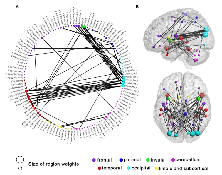
In today’s Research News article “Open monitoring meditation reduces the involvement of brain regions related to memory function.” (See summary below or view the full text of the study at: https://www.ncbi.nlm.nih.gov/pmc/articles/PMC6028418/ ), Fujino and colleagues recruited experienced meditators and scanned their brains for functional connectivity between structures with functional Magnetic Resonance Imaging (f-MRI) while they were engaging in open monitoring meditation, and again when engaging in focused attention meditation. Each f-MRI session was preceded by a week of practicing the appropriate meditation type at home.


They found that both meditation types produced decreased functional connectivity between the striatum, a component of the attention neural network and the posterior cingulate cortex a component of the Default Mode Network that is usually active during mind wandering. These findings suggest that both techniques help keep the mind focused and successfully suppress the mind straying from the task at hand.


The meditation techniques also produced differences in functional connectivity with open monitoring meditation reducing functional connectivity of the ventral striatum with both the visual cortex and retrosplenial cortex while focused attention meditation increasing this connectivity. In some ways this is not surprising as the striatum – visual cortex connection is thought to be involved in intentional focused attention. It would be expected that  focused attention meditation would strengthen this while open monitoring meditation would weaken it. In addition, the visual cortex is thought to be involved in memory. This suggests that open monitoring meditation may reduce the tendency to have memories interjected into the meditation practice.


So, the results are suggestive of similar effects of open monitoring meditation and focused attention meditation on the brain systems maintaining attention and suppressing mind wandering and differing effects on the brain system underlying focused attention and memory. These differing neural changes suggest that the two practices produce different experiential effects on the individual during practice.


“Open monitoring meditation is known to make you more creative. And if you feel as though you are stuck in a rut or as though you need to find alternative solutions to problems, then this can be a very effective techniques to use.

Focused attention meditation, as you know, is all about focusing your mind on one thing at a time, often the breath. This is good for improve focus and concentration, for stopping multitasking, and also for various health reasons.” – Paul Harrison


CMCS – Center for Mindfulness and Contemplative Studies


This and other Contemplative Studies posts are also available on Google+ https://plus.google.com/106784388191201299496/posts and on Twitter @MindfulResearch


Study Summary


Fujino, M., Ueda, Y., Mizuhara, H., Saiki, J., & Nomura, M. (2018). Open monitoring meditation reduces the involvement of brain regions related to memory function. Scientific Reports, 8, 9968. http://doi.org/10.1038/s41598-018-28274-4


Abstract

Mindfulness meditation consists of focused attention meditation (FAM) and open monitoring meditation (OMM), both of which reduce activation of the default mode network (DMN) and mind-wandering. Although it is known that FAM requires intentional focused attention, the mechanisms of OMM remain largely unknown. To investigate this, we examined striatal functional connectivity in 17 experienced meditators (mean total practice hours = 920.6) during pre-resting, meditation, and post-resting states comparing OMM with FAM, using functional magnetic resonance imaging. Both FAM and OMM reduced functional connectivity between the striatum and posterior cingulate cortex, which is a core hub region of the DMN. Furthermore, OMM reduced functional connectivity of the ventral striatum with both the visual cortex related to intentional focused attention in the attentional network and retrosplenial cortex related to memory function in the DMN. In contrast, FAM increased functional connectivity in these regions. Our findings suggest that OMM reduces intentional focused attention and increases detachment from autobiographical memory. This detachment may play an important role in non-judgmental and non-reactive attitude during OMM. These findings provide new insights into the mechanisms underlying the contribution of OMM to well-being and happiness.

https://www.ncbi.nlm.nih.gov/pmc/articles/PMC6028418/


Increase Brain Matter and Memory in Aging with Tai Chi


Increase Brain Matter and Memory in Aging with Tai Chi


By John M. de Castro, Ph.D.


“Tai Chi can be used as exercise to improve the body, as well as reversing the natural tendency for the brain to shrink with age.” – Functional Aging Institute


The aging process involves a systematic progressive decline in every system in the body, the brain included. This includes our mental abilities which decline with age including impairments in memory, attention, and problem solving ability. It is inevitable and cannot be avoided. Using modern neuroimaging techniques, scientists have been able to view the changes that occur in the nervous system with aging. In addition, they have been able to investigate various techniques that might slow the process of neurodegeneration that accompanies normal aging. They’ve found that mindfulness practices reduce the deterioration of the brain that occurs with aging restraining the loss of neural tissue. Indeed, the brains of practitioners of meditation and yoga have been found to degenerate less with aging than non-practitioners.


Hence, there is some hope for age related cognitive decline, as there is evidence that it can be slowed. There are some indications that physical and mental exercise can reduce the rate of cognitive decline and lower the chances of dementia. For example, contemplative practices such as meditationyoga, and Tai Chi and Qigong have all been shown to be beneficial in slowing or delaying physical and mental decline with aging. Mindfulness practices have been shown to improve cognitive processes while gentle mindful exercises such as Tai Chi and Qigong have been shown to slow age related cognitive decline. It would seem reasonable to hypothesize that Tai Chi and Baduanjin practices might decrease age related decreases in cognitive ability and degeneration of the nervous system.


In today’s Research News article “Tai Chi Chuan and Baduanjin increase grey matter volume in older adults: a brain imaging study.” (See summary below or view the full text of the study at: https://www.ncbi.nlm.nih.gov/pmc/articles/PMC5659386/ ), Tao and colleagues recruited healthy sedentary older adults (50-70 years of age) and randomly assigned them to receive either Tai Chi practice, Baduanjin practice (a very similar practice to Tai Chi), or no-treatment. Practice occurred for 12 weeks, 5 days per week, for 1 hour. Before and after training the participants underwent Magnetic Resonance Imaging (MRI) scans of their brains. They were also measured for memory ability.


They found that in comparison to baseline and control participants, the Tai Chi or Baduanjin practice participants had significant increases in the amount of grey matter in the left insula, left putamen, left parahippocampus/hippocampus, left amygdala, and left inferior temporal lobe. Hence, Tai Chi or Baduanjin practice appeared to produce increases in neural tissue. In addition, the Tai Chi or Baduanjin practice participants had significant increases in overall memory ability and visual reproduction memory. These improvements in memory were related to the increases in grey matter, with large increases in neural tissue associated with large improvements in memory. Hence, Tai Chi or Baduanjin practice not only increased neural tissue and memory, but the increases in both changed together in the same direction.


Caution must be taken in interpreting these results as the control condition was inactive. As a result, it cannot be determined if Tai Chi or Baduanjin practice per se or any form of exercise could produce comparable benefits. Further research is needed employing other forms of exercise to compare to the effects of Tai Chi or Baduanjin practice.


Nonetheless, these results are interesting and exciting. They suggest that Tai Chi or Baduanjin practice can reduce or possibly reverse brain degeneration and cognitive decline associated with aging. By engaging in these mindful movement practices aging individuals appear to preserve their brains and their mental ability. In addition, the fact that these practices are safe, convenient, low cost, and social suggests that they can be widely applied to the aging population.


So, increase brain matter and memory in aging with Tai Chi.


“Keep your brain younger longer by adding tai chi to your workout routine.” – Linda Melone


CMCS – Center for Mindfulness and Contemplative Studies


This and other Contemplative Studies posts are also available on Google+ https://plus.google.com/106784388191201299496/posts and on Twitter @MindfulResearch


Study Summary


Tao, J., Liu, J., Liu, W., Huang, J., Xue, X., Chen, X., … Kong, J. (2017). Tai Chi Chuan and Baduanjin increase grey matter volume in older adults: a brain imaging study. Journal of Alzheimer’s Disease : JAD, 60(2), 389–400. http://doi.org/10.3233/JAD-170477


Abstract

The aim of this study is to investigate and compare how 12-weeks of Tai Chi Chuan and Baduanjin exercise can modulate brain structure and memory function in older adults. Magnetic Resonance Imaging(MRI) and memory function measurements (Wechsler Memory Scale-Chinese revised, WMS-CR)were applied at both the beginning and end of the study. Results showed that both Tai Chi Chuan and Baduanjin could significantly increase grey matter volume (GMV) in the insula, medial temporal lobe (MTL), and putamen after 12-weeks of exercise. No significant differences were observed in grey matter volume (GMV) between the Tai Chi Chuan and Baduanjin groups. We also found that compared to healthy controls, Tai Chi Chuan and Baduanjin significantly improved visual reproduction subscores on the WMS-CR. Baduanjin also improved mental control, recognition, touch and comprehension memory subscores of the WMS-CR compared to the control group. Memory quotient (MQ)and visual reproduction subscores were both associated with GMV increases in the putamen and hippocampus. Our results demonstrate the potential of Tai Chi Chuan and Baduanjin exercise for the prevention of memory deficits in older adults.

https://www.ncbi.nlm.nih.gov/pmc/articles/PMC5659386/

Improve Brain Systems Underlying Mental Well-Being with Gratitude Meditation


Improve Brain Systems Underlying Mental Well-Being with Gratitude Meditation

By John M. de Castro, Ph.D.

“benefit of meditation. Or, rather, some ancient benefit that is just now being confirmed with fMRI or EEG. The practice appears to have an amazing variety of neurological benefits – from changes in grey matter volume to reduced activity in the “me” centers of the brain to enhanced connectivity between brain regions.” – Alice Walton

There has accumulated a large amount of research demonstrating that meditation has significant benefits for psychological, physical, and spiritual wellbeing. Its positive effects are so widespread that it is difficult to find any other treatment of any kind with such broad beneficial effects on everything from mood and happiness to severe mental and physical illnesses. This raises the question of how meditation could do this. One possibility is that mindfulness practice results in beneficial changes in the nervous system.

The nervous system is a dynamic entity, constantly changing and adapting to the environment. It will change size, activity, and connectivity in response to experience. These changes in the brain are called neuroplasticity. Over the last decade neuroscience has been studying the effects of contemplative practices on the brain and has identified neuroplastic changes in widespread areas. In other words, mindfulness practice appears to mold and change the brain, producing psychological, physical, and spiritual benefits.

These neuroplastic changes, however, result from the summation of individual changes occurring to the nervous system in real time produced by the immediate behavior. In order to better understand the process by which behavior affects the nervous system, it is important to look initially at the short-term changes produced by behavior. Loving-kindness meditation has been shown to produce improvement in the regulation of emotions and also changes the areas of the nervous system involved in emotion regulation. To better understand the processes involved it is important to look at the short-term effects of focusing on gratitude on the nervous system.

In today’s Research News article “Effects of gratitude meditation on neural network functional connectivity and brain-heart coupling.” See summary below or view the full text of the study at: https://www.ncbi.nlm.nih.gov/pmc/articles/PMC5506019/, Kyeong and colleagues recruited meditation-naïve adults. They were measured before testing for depression, competence, relatedness, and autonomy. The participants laid in a brain scanner and were instructed to think deeply in a gratitude condition and a resentment condition while their heart rate was recorded and functional Magnetic Resonance Imaging (f-MRI) was performed. In the gratitude condition the participants were instructed to think about and visualize their mother for 4 minutes and tell her in their mind how much they love and appreciate her. In the resentment condition the participants were instructed to think about and visualize a person who made them angry. The two conditions were presented in counterbalanced order to two randomly assigned groups.

They found that the heart rate was significantly lower during the gratefulness condition indicating the positive emotional effects of gratitude. In addition, they found that the functional connectivity of brain areas was significantly related to heart rate in the gratefulness condition but not the resentment condition. Hence, experiencing gratefulness connects central and peripheral mechanisms of emotion. They found that the functional connectivity of neural systems were altered during and after the two conditions. But, the gratitude condition altered the functional connectivity of brain areas associated with emotions and motivation.

These results suggest that the immediate focusing on gratitude produces momentary alterations of brain systems associated with the regulation of emotions and self-motivation. When carried out over a period of time this could sum to produce relatively permanent changes in these neural systems. This may be the mechanism by which loving-kindness meditation improves emotional well-being. These results are a useful start at unravelling the processes by which mental contents can produce relatively permanent alterations of the nervous system and thereby produce relatively permanent changes in the individual’s mood and regulation of that mood.

So, improve brain systems underlying mental well-being with gratitude meditation.

“Being distracted exacts a cost on our well-being, If we become more mindful of our everyday activities, we can learn well-being and become happier.” – Ritchie Davidson

CMCS – Center for Mindfulness and Contemplative Studies

This and other Contemplative Studies posts are available at the Contemplative Studies Blog http://contemplative-studies.org/wp/
They are also available on Google+ https://plus.google.com/106784388191201299496/posts and on Twitter @MindfulResearch

Study Summary

Kyeong, S., Kim, J., Kim, D. J., Kim, H. E., & Kim, J.-J. (2017). Effects of gratitude meditation on neural network functional connectivity and brain-heart coupling. Scientific Reports, 7, 5058. http://doi.org/10.1038/s41598-017-05520-9

Abstract
A sense of gratitude is a powerful and positive experience that can promote a happier life, whereas resentment is associated with life dissatisfaction. To explore the effects of gratitude and resentment on mental well-being, we acquired functional magnetic resonance imaging and heart rate (HR) data before, during, and after the gratitude and resentment interventions. Functional connectivity (FC) analysis was conducted to identify the modulatory effects of gratitude on the default mode, emotion, and reward-motivation networks. The average HR was significantly lower during the gratitude intervention than during the resentment intervention. Temporostriatal FC showed a positive correlation with HR during the gratitude intervention, but not during the resentment intervention. Temporostriatal resting-state FC was significantly decreased after the gratitude intervention compared to the resentment intervention. After the gratitude intervention, resting-state FC of the amygdala with the right dorsomedial prefrontal cortex and left dorsal anterior cingulate cortex were positively correlated with anxiety scale and depression scale, respectively. Taken together, our findings shed light on the effect of gratitude meditation on an individual’s mental well-being, and indicate that it may be a means of improving both emotion regulation and self-motivation by modulating resting-state FC in emotion and motivation-related brain regions.
https://www.ncbi.nlm.nih.gov/pmc/articles/PMC5506019/

Protect the Aging Brain with Meditation

Protect the Aging Brain with Meditation


By John M. de Castro, Ph.D.


“long-term engagement in mindfulness meditation may enhance cognitive performance in older adults, and that with persistent practice, these benefits may be sustained. That’s great news for the millions of aging adults working to combat the negative effects of aging on the brain.” B. Grace Bullock


Human life is one of constant change. We revel in our increases in physical and mental capacities during development, but regret their decreases during aging. The aging process involves a systematic progressive decline in every system in the body, the brain included. Starting in the 20s there is a progressive decrease in the volume of the brain as we age.


The nervous system is a dynamic entity, constantly changing and adapting to the environment. It will change size, activity, and connectivity in response to experience. These changes in the brain are called neuroplasticity.  Over the last decade neuroscience has been studying the effects of contemplative practices on the brain and has identified neuroplastic changes in widespread area. and have found that meditation practice appears to mold and change the brain, producing psychological, physical, and spiritual benefits. In addition, they have been able to investigate various techniques that might slow the process of neurodegeneration that accompanies normal aging. They’ve found that mindfulness practices reduce the deterioration of the brain that occurs with aging restraining the loss of neural tissue. Indeed, the brains of practitioners of meditation and yoga have been found to degenerate less with aging than non-practitioners.


In today’s Research News article “Promising Links between Meditation and Reduced (Brain) Aging: An Attempt to Bridge Some Gaps between the Alleged Fountain of Youth and the Youth of the Field.” See summary below or view the full text of the study at: https://www.ncbi.nlm.nih.gov/pmc/articles/PMC5447722/, Kurth and colleagues review and summarize the published research literature on the neuroprotective effects of meditation in the elderly. They discuss the ideas that the aging based deterioration of the brain is due to a number of processes, including changes in the DNA telomeres, inflammation, stress, and neuroplasticity and that meditation appears to effect all of these processes.


There has accumulated evidence that meditation protects against age related decline at the molecular genetic level. As we age the length of a DNA structures called the telomeres progressively shorten. It is thought that the shorter the telomeres get the more difficult it becomes for cells to replicate properly and thus leads to decline. Mindfulness training in general and meditation specifically, has been shown to reduce the shortening of the telomeres with aging. Kurth and colleagues speculate that this is one mechanism by which meditation protects the brain from age related decline.


As we age the natural inflammatory response that normally occurs to protect against infection begins to increase in general and lose its specificity to fighting particular diseases, pathogens, and injuries. It becomes more widespread damaging normal tissues. Mindfulness training in general and meditation specifically has been shown to reduce inflammatory responses. It seems reasonable that this is another mechanism by which meditation protects the body from age related decline.


Stress is present throughout life. But if it is too intense or prolonged the biological responses to stress begin to damage the body. These stress induced changes are similar to age related deterioration. Stress effects may accumulate over time. Hence, the older we get the greater the total stress induced damage. Mindfulness training in general and meditation specifically has been shown to improve emotion regulation and to reduce the physiological and psychological responses to stress. This is hypothesized to be another mechanism by which meditation protects the brain from deterioration with aging.


Neuroplasticity is a change in the size and connectivity of brain structures as they are exercised over a prolonged period of time. Mindfulness training in general and meditation specifically has been shown to produce neuroplastic changes in the brain, increasing the size and connectivity of brain structures. This process would tend to counteract brain degeneration with aging and may be another mechanism by which meditation protects the brain during aging.


Hence there has accumulated evidence that meditation reduces the deterioration of the brain with aging. It appears to do so by altering a number of different mechanisms including changes in the DNA telomeres, inflammation, stress, and neuroplasticity. This protection of the brain may be responsible to the ability of meditation to reduce the decline in mental abilities that occur with aging. This would tend to make aging a more benign process.


So, protect the aging brain with meditation.


We expected rather small and distinct effects located in some of the regions that had previously been associated with meditating. Instead, what we actually observed was a widespread effect of meditation that encompassed regions throughout the entire brain.” – Florian Kurth


CMCS – Center for Mindfulness and Contemplative Studies


This and other Contemplative Studies posts are also available on Google+ https://plus.google.com/106784388191201299496/posts and on Twitter @MindfulResearch


Study Summary


Kurth, F., Cherbuin, N., & Luders, E. (2017). Promising Links between Meditation and Reduced (Brain) Aging: An Attempt to Bridge Some Gaps between the Alleged Fountain of Youth and the Youth of the Field. Frontiers in Psychology, 8, 860. http://doi.org/10.3389/fpsyg.2017.00860


Abstract

Over the last decade, an increasing number of studies has reported a positive impact of meditation on cerebral aging. However, the underlying mechanisms for these seemingly brain-protecting effects are not well-understood. This may be due to the fact, at least partly, that systematic empirical meditation research has emerged only recently as a field of scientific scrutiny. Thus, on the one hand, critical questions remain largely unanswered; and on the other hand, outcomes of existing research require better integration to build a more comprehensive and holistic picture. In this article, we first review theories and mechanisms pertaining to normal (brain) aging, specifically focusing on telomeres, inflammation, stress regulation, and macroscopic brain anatomy. Then, we summarize existing research integrating the developing evidence suggesting that meditation exerts positive effects on (brain) aging, while carefully discussing possible mechanisms through which these effects may be mediated.

https://www.ncbi.nlm.nih.gov/pmc/articles/PMC5447722/

Change the Brain with Mindfulness

Change the Brain with Mindfulness


By John M. de Castro, Ph.D.


“The picture we have is that mindfulness practice increases one’s ability to recruit higher order, pre-frontal cortex regions in order to down-regulate lower-order brain activity. In other words, our more primal responses to stress seem to be superseded by more thoughtful ones.” – Tom Ireland


There has accumulated a large amount of research demonstrating that meditation has significant benefits for psychological, physical, and spiritual wellbeing. Its positive effects are so widespread that it is difficult to find any other treatment of any kind with such broad beneficial effects on everything from mood and happiness to severe mental and physical illnesses. This raises the question of how meditation could do this. One possibility is that mindfulness practice results in beneficial changes in the nervous system.


The nervous system is a dynamic entity, constantly changing and adapting to the environment. It will change size, activity, and connectivity in response to experience. These changes in the brain are called neuroplasticity. Over the last decade neuroscience has been studying the effects of contemplative practices on the brain and has identified neuroplastic changes in widespread areas. In other words, mindfulness practice appears to mold and change the brain, producing psychological, physical, and spiritual benefits.


The results of the research concerning the changes in the brain that occur with mindfulness practice have not presented a consistent picture. One issue may be the way that mindfulness is measured. This issue was explored in today’s Research News article “A distinction between two instruments measuring dispositional mindfulness and the correlations between those measurements and the neuroanatomical structure.” See summary below or view the full text of the study at: https://www.ncbi.nlm.nih.gov/pmc/articles/PMC5524689/, Zhuang and colleagues compared Magnetic Resonance Images (MRIs), depression, and emotion regulation, between college students who had been measured for mindfulness with Mindful Attention Awareness Scale (MAAS) to those measured with the Five Factor Mindfulness Questionnaire (FFMQ).


They found that higher MAAS scores and FFMQ Describing, Acting with Awareness, and Non-judging scores the lower levels of depression. In addition, higher FFMQ Describing scores were significantly associated with higher emotion regulation. In regard to the brain scans they found that the higher the MAAS score the greater the size of the Precuneus area of the cortex. Mediation analysis demonstrated that the higher the volume of the precuneus cortical region the higher the MAAS score which in turn was associated with lower depression. So, mindfulness as measured by the MAAS was associated greater volume of the precuneus and was responsible for the relationship of the Precuneus volume with depression.


In regard to mindfulness measured with the FFMQ and the brain scans they found that the larger the size of the Superior Prefrontal Cortex, the higher the Describing and Non-judging facets and the lower the Non-reacting facet. In addition, the larger the size of the dorsolateral prefrontal cortex and the inferior parietal cortex the higher the Describing FFMQ facet. Mediation analysis demonstrated that the higher the volume of the Superior Prefrontal Cortex the higher the Describing FFMQ facet which in turn predicted higher emotion regulation. So, mindfulness as measured with the Describing FFMQ facet was associated greater size of the Superior Prefrontal Cortex and was responsible for the relationship of the Superior Prefrontal Cortex size with emotion regulation.


These results are interesting and suggest that the kind of relationship observed between changes in the brain and mindfulness is affected by the way mindfulness is measured. This could account for some of the conflicting findings in the published research. Also, since the Precuneus is associated with awareness of self, the results suggest that mindfulness as measured by the MAAS mainly measures self-awareness while since the inferior parietal cortex is also associated with awareness of self, the results suggest that mindfulness as measured by the Describing FFMQ facet also measures self-awareness. But the FFMQ mindfulness measure goes further and also documents other abilities. Since, the Prefrontal cortex is associated with attention control and emotion regulation, the results suggest that mindfulness as measured by the Describing and Non-reacting FFMQ facets also measure attention control and emotion regulation.


Clearly, mindfulness is associated with different sizes of areas in the brain’s cortical regions. But, even though the brain is different with mindfulness, the types of differences observed depends upon how mindfulness is measured. The Five Factor Mindfulness Questionnaire (FFMQ) measure appears to be superior to the Mindful Attention Awareness Scale (MAAS) as it breaks mindfulness down into component parts providing greater refinement in observed brain changes. These results will be helpful in future research unravelling the relationship of mindfulness to the characteristics of the nervous system.


The practice of mindfulness can train our brains to have a new default. Instead of automatically falling into the stream of past or future rumination that ignites the depression loop, mindfulness draws our attention to the present moment. As we practice mindfulness, we actually start wiring neurons that balance the brain in a way that is naturally an antidepressant. “ – Debbie Hampton


CMCS – Center for Mindfulness and Contemplative Studies


This and other Contemplative Studies posts are also available on Google+ https://plus.google.com/106784388191201299496/posts and on Twitter @MindfulResearch


Study Summary


Zhuang, K., Bi, M., Li, Y., Xia, Y., Guo, X., Chen, Q., … Qiu, J. (2017). A distinction between two instruments measuring dispositional mindfulness and the correlations between those measurements and the neuroanatomical structure. Scientific Reports, 7, 6252. http://doi.org/10.1038/s41598-017-06599-w


Abstract

The most widely used measurements of mindfulness are the Mindful Attention Awareness Scale (MAAS) and the Five Facet Mindfulness Questionnaire (FFMQ). However, controversies exist regarding the application of these scales. Additionally, the neural mechanisms of dispositional mindfulness have become a topic of interest. In the current study, we used surface-based methodology to identify the brain regions underlying individual differences in dispositional mindfulness in a large non-clinical sample and compared the two instruments for measuring the dispositional mindfulness. The results indicated that the MAAS scores were significantly associated with increased grey matter volumes in the right precuneus and the significant association between the precuneus and depression symptomatology was mediated by MAAS scores. Regarding the FFMQ, the Describing, Nonjudging, and Nonreactivity facets were selectively associated with the cortical volume, thickness and surface area of multiple prefrontal regions as well as the inferior parietal lobule. Importantly, Describing mediated the association between the dorsolateral PFC volume and the cognitive reappraisal strategies of emotion regulation. These results suggested that the MAAS were mainly associated with self-awareness, while the FFMQ facets were selectively involved in emotion regulation, attention control and self-awareness. Therefore, this study characterized the differences in inter-individual variability between the two typical measurements of dispositional mindfulness and the correlations between those measurements and imaging analyses.

https://www.ncbi.nlm.nih.gov/pmc/articles/PMC5524689/

Alter Brain Networks and Cognitive Ability with Tai Chi

Alter Brain Networks and Cognitive Ability with Tai Chi


By John M. de Castro, Ph.D.


” Another great benefit of Tai Chi is that it’s accessible to people of all ages and fitness abilities. It’s the focus on the subtle movements that exercise the brain and boost cognitive abilities.” – Karl Romain


Tai Chi is an ancient Chinese practice involving mindfulness and gentle movements. It is easy to learn, safe, and gentle. Tai Chi has been practiced for thousands of years with benefits for health and longevityTai Chi training is designed to enhance function and regulate the activities of the body through controlled breathing, mindful concentration, and gentle movements. Only recently though have the effects of this practice been scrutinized with empirical research. This research has found that it is effective for an array of physical and psychological issues. It appears to strengthen the immune systemreduce inflammation and increase the number of cancer killing cells in the bloodstream, improve cardiovascular healthreduce arthritis painimprove balance and reduce falls. It also appears to improve attentional ability and relieve depression.


The nervous system is a dynamic entity, constantly changing and adapting to the environment. It will change size, activity, and connectivity in response to experience. These changes in the brain are called neuroplasticity. Over the last decade neuroscience has been studying the effects of contemplative practices on the brain and has identified neuroplastic changes in widespread areas. In other words, mindfulness practice appears to mold and change the brain, producing psychological, physical, and spiritual benefits. Hence, it would appear likely that Tai Chi practice may alter the brain networks underlying mindfulness.


In today’s Research News article “Mind-Body Practice Changes Fractional Amplitude of Low Frequency Fluctuations in Intrinsic Control Networks.” See summary below or view the full text of the study at: http://journal.frontiersin.org/article/10.3389/fpsyg.2017.01049/full?utm_source=F-AAE&utm_medium=EMLF&utm_campaign=MRK_325025_69_Psycho_20170711_arts_A

Wei and colleagues recruited healthy long-term practitioners of Tai Chi (average 14 years of practice) and age, gender, and education matched non-practitioners. All participants performed a flanker task, a measure of executive cognitive function, in which the participant had to respond to the direction of an arrow surrounded by distracting arrows. They found that the longer the practitioners had been practicing Tai Chi the faster they responded in the flanker, executive function, task. This suggests that Tai Chi practice enhances cognitive function.


The participants then underwent functional Magnetic Resonance Imaging (fMRI) brain scanning. The brain scans revealed that the Tai Chi practitioners had a significant reduction in amplitude of low frequency fluctuations in the brain areas called the Default Mode Network (DMN) which underlies mind wandering and self-referential thinking. There was also a significant reduction in amplitude of low frequency fluctuations in the brain areas called the frontoparietal network (FPN) and the dorsal prefrontal-angular network which are associated with cognitive control and executive function. These changes in the low frequency fluctuations suggest that Tai Chi practice produces changes in these networks increasing their functional connectivity.


These results are very interesting and suggest that Tai Chi practice can produce changes in the brain improving the connectivities within large-scale neural systems. At least in terms of the frontoparietal network (FPN) and the dorsal prefrontal-angular network these changes may underlie the ability of Tai Chi practice to improve cognitive control and executive function while the changes in the Default Mode Network (DMN) may underlie the ability of Tai Chi practice to reduce mind wandering and self-referential thinking. Hence, Tai Chi practice alters the nervious system and produces very beneficial effects


So, alter brain networks and cognitive ability with Tai Chi.


“Neuroplasticity may sound like an out of this world term to a normal Tai Chi practitioner but to those who pursue the path of the mind, Warriors of Intention, then this is the proven way of enabling our mind to permanently rewire the way we move.” – Mushin


CMCS – Center for Mindfulness and Contemplative Studies


This and other Contemplative Studies posts are also available on Google+ https://plus.google.com/106784388191201299496/posts and on Twitter @MindfulResearch


Study Summary


Wei G-X, Gong Z-Q, Yang Z and Zuo X-N (2017) Mind-Body Practice Changes Fractional Amplitude of Low Frequency Fluctuations in Intrinsic Control Networks. Front. Psychol. 8:1049. doi: 10.3389/fpsyg.2017.01049


Abstract

Cognitive control impairment is a typical symptom largely reported in populations with neurological disorders. Previous studies have provided evidence about the changes in cognitive control induced by mind-body training. However, the neural correlates underlying the effect of extensive mind-body practice on cognitive control remain largely unknown. Using resting-state functional magnetic resonance imaging, we characterized dynamic fluctuations in large-scale intrinsic connectivity networks associated with mind-body practice, and examined their differences between healthy controls and Tai Chi Chuan (TCC) practitioners. Compared with a control group, the TCC group revealed significantly decreased fractional Amplitude of Low Frequency Fluctuations (fALFF) in the bilateral frontoparietal network, default mode network and dorsal prefrontal-angular gyri network. Furthermore, we detected a significant association between mind-body practice experience and fALFF in the default mode network, as well as an association between cognitive control performance and fALFF of the frontoparietal network. This provides the first evidence of large-scale functional connectivity in brain networks associated with mind-body practice, shedding light on the neural network changes that accompany intensive mind-body training. It also highlights the functionally plastic role of the frontoparietal network in the context of the “immune system” of mental health recently developed in relation to flexible hub theory.

http://journal.frontiersin.org/article/10.3389/fpsyg.2017.01049/full?utm_source=F-AAE&utm_medium=EMLF&utm_campaign=MRK_325025_69_Psycho_20170711_arts_A

Meditation Practice Alters Brain Networks in the Elderly

Meditation Practice Alters Brain Networks in the Elderly


By John M. de Castro, Ph.D.


“A growing body of research supports the immediate benefits of meditation, such as reduced stress and anxiety levels, lower blood pressure, and enhanced happiness. Studies on mindfulness interventions show these effects are common in as few as eight weeks. While these initial perks may be reason enough for us to practice, meditation’s positive impact appears to be even more far-reaching, potentially adding years to our lives and improving cognitive function well into old age.” – Rina Deshpande


Human life is one of constant change. We revel in our increases in physical and mental capacities during development, but regret their decreases during aging. The aging process involves a systematic progressive decline in every system in the body, the brain included. This includes our mental abilities which decline with age including impairments in memory, attention, and problem solving ability. It is inevitable and cannot be avoided.


The nervous system is a dynamic entity, constantly changing and adapting to the environment. It will change size, activity, and connectivity in response to experience. These changes in the brain are called neuroplasticity. Over the last decade neuroscience has been studying the effects of contemplative practices on the brain and has identified neuroplastic changes in widespread area. and have found that meditation practice appears to mold and change the brain, producing psychological, physical, and spiritual benefits. In addition, they have been able to investigate various techniques that might slow the process of neurodegeneration that accompanies normal aging. They’ve found that mindfulness practices reduce the deterioration of the brain that occurs with aging restraining the loss of neural tissue. Indeed, the brains of practitioners of meditation and yoga have been found to degenerate less with aging than non-practitioners.


The changes in the aging brain produced by meditation are not a simple increases in tissue throughout the brain but rather increases in some area and systems and decreases in others. In today’s Research News article “A longitudinal study of the effect of short-term meditation training on functional network organization of the aging brain.” See summary below or view the full text of the study at: https://www.ncbi.nlm.nih.gov/pmc/articles/PMC5428857/, Cotier and colleagues studied the neuroplastic changes in brain systems produced by meditation in the elderly. They recruited health elderly participants (average age of years) and randomly assigned them to receive either 8 weeks of meditation or relaxation training. Training occurred in 22 90-minute group classes. “Meditation participants were taught to cultivate mindfulness through paying attention to the surrounding sounds and one’s own breathing, feelings and sensations on the present moment.” “Relaxation participants were taught diaphragmatic breathing, progressive muscle relaxation and imagery relaxation techniques aimed at enhancing body awareness and reducing body tension.”


Before and after training the participants underwent functional Magnetic Resonance Imaging (fMRI) scanning of their brains. They found that after 8-weeks of meditation practice there was reduced functional connectivity in the default mode network (DMN), salience network (SN), and somatomotor network (SMN) while after relaxation training these same systems showed increased functional connectivity. Also, in the meditation group “there was decreased connectivity strength between the DMN and other modules.”


These are very interesting results as the Default Mode Network (DMN) becomes active during wind wandering and relatively quiet during focused on task behavior. It is involved when we are engaged in internally focused tasks such as recalling deeply personal memories, daydreaming, sleeping, imagining the future and trying to take the perspective of others. The DMN involves neural structures including the medial prefrontal cortex, anterior and posterior cingulate cortices, precuneus, inferior parietal cortex, and lateral temporal cortex. These areas of the DMN are functionally connected, such that they are simultaneously active during mind wandering.


Hence the current results of reduced functional connectivity in the DMN and between the DMN and other neural systems suggests that the meditation practice resulted in changes in the brain reflective of decreased mind wandering and increased focused attention. These changes can be helpful to the elderly who demonstrate increasing difficulties with attention. These changes may underlie the ability of meditation practice to reduce cognitive decline in the elderly. Hence, they suggest that meditation practice by the elderly can change their nervous systems in beneficial ways, decreasing the tendency for the mind to wander away from the task at hand and improving attentional ability and thereby reducing the deterioration of thought processes.


So, alter brain networks in the elderly with meditation practice.


“at a time when Americans are living longer than ever before but experiencing higher rates of Alzheimer’s and other neurodegenerative disorders, the research is an important reminder that mindfulness can exert a long-lasting influence on brain health.”Carolyn Gregoire


CMCS – Center for Mindfulness and Contemplative Studies


This and other Contemplative Studies posts are also available on Google+ https://plus.google.com/106784388191201299496/posts and on Twitter @MindfulResearch


Study Summary


Cotier, F. A., Zhang, R., & Lee, T. M. C. (2017). A longitudinal study of the effect of short-term meditation training on functional network organization of the aging brain. Scientific Reports, 7, 598. http://doi.org/10.1038/s41598-017-00678-8


Abstract

The beneficial effects of meditation on preserving age-related changes in cognitive functioning are well established. Yet, the neural underpinnings of these positive effects have not been fully unveiled. This study employed a prospective longitudinal design, and graph-based analysis, to study how an eight-week meditation training vs. relaxation training shaped network configuration at global, intermediate, and local levels using graph theory in the elderly. At the intermediate level, meditation training lead to decreased intra-connectivity in the default mode network (DMN), salience network (SAN) and somatomotor network (SMN) modules post training. Also, there was decreased connectivity strength between the DMN and other modules. At a local level, meditation training lowered nodal strength in the left posterior cingulate gryus, bilateral paracentral lobule, and middle cingulate gyrus. According to previous literature, the direction of these changes is consistent with a movement towards a more self-detached viewpoint, as well as more efficient processing. Furthermore, our findings highlight the importance of considering brain network changes across organizational levels, as well as the pace at which these changes may occur. Overall, this study provides further support for short-term meditation as a potentially beneficial method of mental training for the elderly that warrants further investigation.

https://www.ncbi.nlm.nih.gov/pmc/articles/PMC5428857/

Change the Brain with Brief Mindfulness Training

Change the Brain with Brief Mindfulness Training


By John M. de Castro, Ph.D.


“We can intentionally shape the direction of plasticity changes in our brain. By focusing on wholesome thoughts, for example, and directing our intentions in those ways, we can potentially influence the plasticity of our brains and shape them in ways that can be beneficial. That leads us to the inevitable conclusion that qualities like warm-heartedness and well-being should best be regarded as skills.” – Richie Davidson


There has accumulated a large amount of research demonstrating that meditation has significant benefits for psychological, physical, and spiritual wellbeing. Its positive effects are so widespread that it is difficult to find any other treatment of any kind with such broad beneficial effects. They range from emotion regulation, attention, cognitive performance and happiness to severe mental and physical illnesses. These effects appear to be relatively permanent which suggests that mindfulness meditation produces some relatively permanent change in the brain.


The nervous system is a dynamic entity, constantly changing and adapting to the environment. It will change size, activity, and connectivity in response to experience. For example, the brain area that controls the right index finger has been found to be larger in blind subjects who use braille than in sighted individuals.  Similarly, cab drivers in London who navigate the twisting streets of the city, have a larger hippocampus, which is involved in spatial navigation, than predefined route bus drivers. These changes in the brain are called neuroplasticity. Over the last decade neuroscience has been studying the effects of contemplative practices on the brain and has identified neuroplastic changes in widespread areas. In other words, meditation practice appears to mold and change the brain, producing psychological, physical, and spiritual benefits.


Although the effectiveness of meditation in producing psychological and physical benefits and in producing neuroplastic changes to the brain, the needed dose is not known. In other words, there is a need to investigate the effectiveness of different amounts of meditation practice and exactly what changes they produce in the brain. In today’s Research News article “Brief Mental Training Reorganizes Large-Scale Brain Networks.” See summary below or view the full text of the study at: https://www.ncbi.nlm.nih.gov/pmc/articles/PMC5328965/

Tang and colleagues investigate the effects of a brief mindfulness meditation training, 10 30-minute sessions over 2 weeks, on functional connections between brain structures. The meditation training was called Integrative Body–Mind Training (IBMT) and includes body relaxation, mental imagery and open monitoring mindfulness meditation.


They recruited meditation naive male and female undergraduate students and collected functional Magnetic Resonance Imaging (fMRI) scans before and after training and compared resting functional connectivity after training to that observed at baseline. They found 105 different connectivity changes in the brains following mindfulness meditation practice. Functional connectivity was increased between the Occipital Cortex and a wide range of other areas, particularly in the Temporal Cortex, mainly the superior temporal gyrus and its pole, and the insula, and also with the frontal cortex, mainly the frontal operculum.


The study demonstrated that even a brief mindfulness meditation training of only 10 total hours of practice produces widespread changes in the nervous system. This is remarkable that such a small amount of training could produce such profound changes. This testifies to the power of mindfulness training to alter how our physiology processes experience. It is curious that the Occipital Cortex was found to be so involved. Occipital Cortex is involved in visual processing but Integrative Body–Mind Training (IBMT) is practiced with eyes closed. It is possible that the mental imagery was responsible for this involvement. But, it will take more research to understand the nature of the observed changes. Regardless it is clear that major changes in brain connectivity are produced even by brief mindfulness meditation practice.


So, change the brain with brief mindfulness training.


“Now, as the popularity of mindfulness grows, brain imaging techniques are revealing that this ancient practice can profoundly change the way different regions of the brain communicate with each other – and therefore how we think – permanently.” – Tom Ireland


CMCS – Center for Mindfulness and Contemplative Studies


This and other Contemplative Studies posts are also available on Google+ https://plus.google.com/106784388191201299496/posts and on Twitter @MindfulResearch


Study Summary

Tang, Y.-Y., Tang, Y., Tang, R., & Lewis-Peacock, J. A. (2017). Brief Mental Training Reorganizes Large-Scale Brain Networks. Frontiers in Systems Neuroscience, 11, 6. http://doi.org/10.3389/fnsys.2017.00006


Abstract

Emerging evidences have shown that one form of mental training—mindfulness meditation, can improve attention, emotion regulation and cognitive performance through changing brain activity and structural connectivity. However, whether and how the short-term mindfulness meditation alters large-scale brain networks are not well understood. Here, we applied a novel data-driven technique, the multivariate pattern analysis (MVPA) to resting-state fMRI (rsfMRI) data to identify changes in brain activity patterns and assess the neural mechanisms induced by a brief mindfulness training—integrative body–mind training (IBMT), which was previously reported in our series of randomized studies. Whole brain rsfMRI was performed on an undergraduate group who received 2 weeks of IBMT with 30 min per session (5 h training in total). Classifiers were trained on measures of functional connectivity in this fMRI data, and they were able to reliably differentiate (with 72% accuracy) patterns of connectivity from before vs. after the IBMT training. After training, an increase in positive functional connections (60 connections) were detected, primarily involving bilateral superior/middle occipital gyrus, bilateral frontale operculum, bilateral superior temporal gyrus, right superior temporal pole, bilateral insula, caudate and cerebellum. These results suggest that brief mental training alters the functional connectivity of large-scale brain networks at rest that may involve a portion of the neural circuitry supporting attention, cognitive and affective processing, awareness and sensory integration and reward processing.

https://www.ncbi.nlm.nih.gov/pmc/articles/PMC5328965/


Change the Brain with Different Meditation Practices

Change the Brain with Different Meditation Practices


By John M. de Castro, Ph.D.


“Most people tend to believe that all types of meditations are the same. It is common to hear about the benefits of “meditation,” but most people don’t know that there are different benefits to be obtained based specifically on the type of meditation practice pursued.“ – Mental Health Blog


Meditation training has been shown to improve health and well-being. It has also been found to be effective for a large array of medical and psychiatric conditions, either stand-alone or in combination with more traditional therapies. As a result, meditation training has been called the third wave of therapies. One problem with understanding meditation effects is that there are, a wide variety of meditation techniques and it is not known which type or which component produce which effects.


There are a number of different types of meditation. Many can be characterized on a continuum with the degree of attentional focus. In focused attention meditation, the individual practices paying attention to a single meditation object, learns to filter out distracting stimuli, including thoughts, and learns to stay focused on the present moment, filtering out thoughts centered around the past or future. On the other hand, in open monitoring meditation, the individual opens up awareness to everything that’s being experienced regardless of its origin. These include bodily sensations, external stimuli, and even thoughts. The meditator just observes these stimuli and lets them arise, and fall away without paying them any further attention.


One potential method to discern the different effects of these differing meditation techniques is to observe the effects of these techniques on the nervous system. There is evidence that meditation alters the brain. It can produce relatively permanent changes to the nervous system, increasing the activity, size, and connectivity of some structures while decreasing it for others in a process known as neuroplasticity. A common method to study the activity of the nervous system is to measure the electrical signal at the scalp above brain regions. Changes in this activity are measurable with mindfulness training.


In today’s Research News article “Increased Gamma Brainwave Amplitude Compared to Control in Three Different Meditation Traditions.” See summary below or view the full text of the study at:

https://www.ncbi.nlm.nih.gov/pmc/articles/PMC5261734/

Braboszcz and colleagues investigate the effects of three different kinds of meditation that differ on the degree of attentional focus on the electrical activity of the brain (EEG) under different mental states. They investigated the effects of a focused meditation employing mantra repetition (Himalayan Yoga), an open monitoring meditation focusing on body sensations (Vipassana) and a complete open awareness meditation technique (“Shoonya” meditation). They recruited experienced meditators who practiced one of the three techniques and also a group of comparable participants with no meditation experience. The Electroencephalogram (EEG) of the participants was measured during 20 minutes of meditation and 20 minutes of instructed mind wandering.


They found that regardless of the meditation or mind wandering condition the three meditation groups in comparison to the non-meditators had significantly larger amounts of high frequency waves (gamma – 60-110 cycles per second, Hz.) in the EEG. They also found that the amount of gamma activity was associated with the amount of meditative experience of the practitioners, with the more the experience, the greater the gamma activity. In addition, they found that the Vipassana meditation practitioners had significantly larger amounts of low frequency waves (alpha – 8-11 cycles per second, Hz.) than the other meditation groups or the controls regardless of condition.


The fact that the differences in the gamma activity in the EEG of the three groups of meditators compared to controls were present regardless of the meditation or the mind wandering condition, suggests that the increased gamma activity results from relatively permanent changes in the brain produced by the meditation training, neuroplasticity. Gamma activity is generally associated with an overall attentive state. Hence, the results suggest that meditation practice, regardless of type, strengthens attentiveness. This is compatible with the findings that meditation training improves attentional ability.


The fact that the differences in the alpha activity in the EEG of the Vipassana meditation practitioners compared to controls and the other two meditation groups were present regardless of the meditation or the mind wandering condition, suggests that the increased alpha activity also results from relatively permanent changes in the brain neuroplasticity. It is interesting that this group of meditators differed from the other groups in alpha activity. High levels of alpha waves have been associated with selective attention where the individual ignores most stimuli to focus on only a specific set of stimuli. Hence, this suggests that the Vipassana practice, which focuses on internal sensations of the body, may be superior to the other meditation techniques in developing selective attentional ability.


Regardless, the results suggest that practicing meditation produces relatively permanent changes in the brain that results in improved attentional ability and focusing on internal sensations during the meditation produces relatively permanent changes in the brain that results in improved selective attentional ability.


“There are many systems of meditation that widely differ from one another in their procedures, contents, objects, beliefs, and goals.  Given these differences, it is not surprising that research has shown they have different subjective and objective effects.” – David Johnson


CMCS – Center for Mindfulness and Contemplative Studies


This and other Contemplative Studies posts are also available on Google+ https://plus.google.com/106784388191201299496/posts and on Twitter @MindfulResearch


Study Summary

Braboszcz, C., Cahn, B. R., Levy, J., Fernandez, M., & Delorme, A. (2017). Increased Gamma Brainwave Amplitude Compared to Control in Three Different Meditation Traditions. PLoS ONE, 12(1), e0170647. http://doi.org/10.1371/journal.pone.0170647


Abstract

Despite decades of research, effects of different types of meditation on electroencephalographic (EEG) activity are still being defined. We compared practitioners of three different meditation traditions (Vipassana, Himalayan Yoga and Isha Shoonya) with a control group during a meditative and instructed mind-wandering (IMW) block. All meditators showed higher parieto-occipital 60–110 Hz gamma amplitude than control subjects as a trait effect observed during meditation and when considering meditation and IMW periods together. Moreover, this gamma power was positively correlated with participants meditation experience. Independent component analysis was used to show that gamma activity did not originate in eye or muscle artifacts. In addition, we observed higher 7–11 Hz alpha activity in the Vipassana group compared to all the other groups during both meditation and instructed mind wandering and lower 10–11 Hz activity in the Himalayan yoga group during meditation only. We showed that meditation practice is correlated to changes in the EEG gamma frequency range that are common to a variety of meditation practices.

https://www.ncbi.nlm.nih.gov/pmc/articles/PMC5261734/


Change the Brain to Deal with Uncomfortable Sensations with Mindfulness

Change the Brain to Deal with Uncomfortable Sensations with Mindfulness


By John M. de Castro, Ph.D.


“Now, as the popularity of mindfulness grows, brain imaging techniques are revealing that this ancient practice can profoundly change the way different regions of the brain communicate with each other – and therefore how we think – permanently.” – Tom Ireland


The nervous system is a dynamic entity, constantly changing and adapting to the environment. It will change size, activity, and connectivity in response to experience. For example, the brain area that controls the right index finger has been found to be larger in blind subjects who use braille than in sighted individuals.  Similarly, cab drivers in London who navigate the twisting streets of the city, have a larger hippocampus, which is involved in spatial navigation, than predefined route bus drivers. These changes in the brain are called neuroplasticity. Over the last decade neuroscience has been studying the effects of contemplative practices on the brain and has identified neuroplastic changes in widespread areas. In other words, mindfulness practice appears to produce relatively permanent changes in the brain, producing psychological, physical, and spiritual benefits.


Dealing with aversive or painful stimuli can be stressful and difficult. There are, however, methods that can improve the individual’s ability to effectively cope with them. Indeed, mindfulness training has been shown to reduce the experience of and response to aversive stimuli and to reduce the physiological and psychological responses to stress. There are indications that mindfulness training may do so by altering the nervous system. The brain regions of the insula and the anterior cingulate cortex have been shown to be involved in interoceptive awareness, that is the conscious appreciation of the internal state of the body. Hence, these structures would be involved in the processing of aversive and painful stimuli. It would seem reasonable, then, to theorize that mindfulness training improves coping with the pain and stress produced by aversive stimuli by changing the activation of the insula and the anterior cingulate cortex.


In today’s Research News article “Mindfulness-based training attenuates insula response to an aversive interoceptive challenge.” See summary below or view the full text of the study at:

https://www.ncbi.nlm.nih.gov/pmc/articles/PMC4692309/

Haase and colleagues recruited U.S. Marines who were undergoing pre-deployment training. They were randomly assigned to receive either the usual training or the training plus 8 weeks of mindfulness training occurring in weekly 2-hour sessions. They were also encouraged to practice 30 minutes per day by themselves. Before and after training they were measured for mindfulness, response to stressful experiences, and sleep quality. In addition, both before and after training the Marines completed a vigilance task while their brains underwent Magnetic Resonance Imaging (MRI scans). While they were undergoing scanning, periodically they had their breathing restricted by increasing the load on the lungs to inhale for a number of 1-minute periods. This produced oxygen restriction that was aversive and stressful. The participants rated how aversive the breathing restriction was.


They found that the breathing restriction was indeed aversive for both groups and there was no effect of mindfulness training on the aversiveness of the restriction. The mindfulness trained Marines, however, had significantly reduced neural responses from the insula and the anterior cingulate cortex after mindfulness training, while the control group did not. Hence, although mindfulness training did not change the perceived aversiveness of the breathing restriction, it did reduce the response of the brain areas responsible for interoceptive awareness.


These results suggest that mindfulness training produced neuroplastic changes in the nervous system, altering the brain areas that are responsible for reacting and consciously appreciating aversive conditions.  It has been previously demonstrated that mindfulness training reduces the experience of, and response to aversive stimuli and stress. Hence, the present findings suggest that neuroplastic alterations to the insula and the anterior cingulate cortex produced by mindfulness training may underlie the improved ability to cope with aversive stimuli.


So, change the brain to deal with uncomfortable sensations with mindfulness.


“Neuroscientists have also shown that practicing mindfulness affects brain areas related to perception, body awareness, pain tolerance, emotion regulation, introspection, complex thinking, and sense of self. While more research is needed to document these changes over time and to understand underlying mechanisms, the converging evidence is compelling.” – Christina Congleton


CMCS – Center for Mindfulness and Contemplative Studies


This and other Contemplative Studies posts are also available on Google+ https://plus.google.com/106784388191201299496/posts and on Twitter @MindfulResearch


Study Summary

Haase, L., Thom, N. J., Shukla, A., Davenport, P. W., Simmons, A. N., Stanley, E. A., … Johnson, D. C. (2016). Mindfulness-based training attenuates insula response to an aversive interoceptive challenge. Social Cognitive and Affective Neuroscience, 11(1), 182–190. http://doi.org/10.1093/scan/nsu042


Abstract

Neuroimaging studies of mindfulness training (MT) modulate anterior cingulate cortex (ACC) and insula among other brain regions, which are important for attentional control, emotional regulation and interoception. Inspiratory breathing load (IBL) is an experimental approach to examine how an individual responds to an aversive stimulus. Military personnel are at increased risk for cognitive, emotional and physiological compromise as a consequence of prolonged exposure to stressful environments and, therefore, may benefit from MT. This study investigated whether MT modulates neural processing of interoceptive distress in infantry marines scheduled to undergo pre-deployment training and deployment to Afghanistan. Marines were divided into two groups: individuals who received training as usual (control) and individuals who received an additional 20-h mindfulness-based mind fitness training (MMFT). All subjects completed an IBL task during functional magnetic resonance imaging at baseline and post-MMFT training. Marines who underwent MMFT relative to controls demonstrated a significant attenuation of right anterior insula and ACC during the experience of loaded breathing. These results support the hypothesis that MT changes brain activation such that individuals process more effectively an aversive interoceptive stimulus. Thus, MT may serve as a training technique to modulate the brain’s response to negative interoceptive stimuli, which may help to improve resilience.

https://www.ncbi.nlm.nih.gov/pmc/articles/PMC4692309/