Improve Brain Processing of Awareness and Emotions with Mindfulness

Improve Brain Processing of Awareness and Emotions with Mindfulness

By John M. de Castro, Ph.D.


Evidence suggests that particular areas of the brain may either shrink or grow in response to regular mindfulness practice.” – Meera Joshi


There has accumulated a large amount of research demonstrating that meditation practice has significant benefits for psychological, physical, and spiritual wellbeing. One way that meditation practices may produce these benefits is by altering the brain. The nervous system is a dynamic entity, constantly changing and adapting to the environment. It will change size, activity, and connectivity in response to experience. These changes in the brain are called neuroplasticity.


Neuroscience has established that contemplative practices produce neuroplastic changes in widespread areas of the nervous system. In other words, mindfulness practices appears to mold and change the brain, producing psychological, physical, and spiritual benefits. One mindfulness therapeutic technique, Mindfulness-Based Stress Reduction (MBSR), has been commonly applied to the treatment of multiple physical and psychological conditions. The MBSR practice consists of discussion, meditation, yoga, and body scan practices. It is important to understand what are the exact changes in the brain that are produced by the MBSR training.


In today’s Research News article “Alterations of Regional Homogeneity and Functional Connectivity Following Short-Term Mindfulness Meditation in Healthy Volunteers.” (See summary below or view the full text of the study at: https://www.ncbi.nlm.nih.gov/pmc/articles/PMC6813410/), Xiao and colleagues recruited meditation naive healthy adults who were participating in an MBSR training and a matched group of no-treatment control participants. The MBSR program met for 2 hours once a week for 8 weeks along with daily home practice. They were measured before and after training for mindfulness and positive and negative emotions. In addition, the participants’ brains were scanned with a functional magnetic resonance imaging (fMRI) technique.


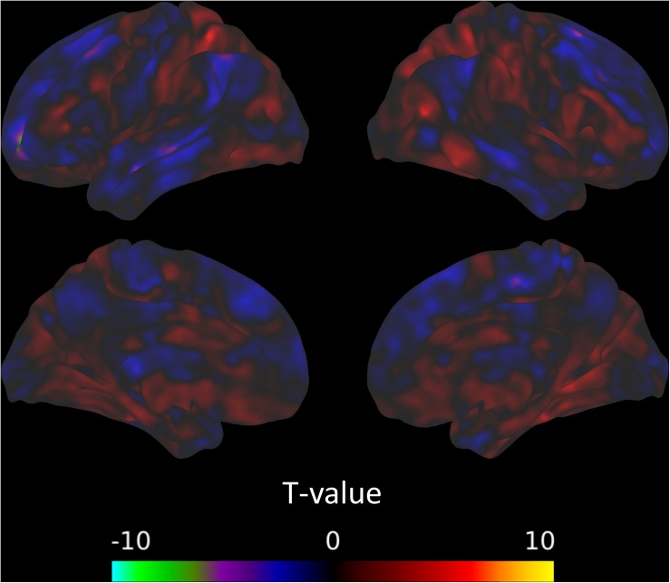
They found that in comparison to baseline and the no-treatment controls, the participants in the Mindfulness-Based Stress Reduction (MBSR) training had significant increases in mindfulness and significant decreases in negative emotions. They also found that following MBSR training there were significant increases in the synchronization of activities in the parietal lobe and significant decreases in the synchronization of activities in the posterior cingulate cortex, precuneus, and cuneus, and increased functional connectivity in the superior parietal lobule and postcentral gyrus and the median cingulate cortex, insula and precentral gyrus.


The findings that Mindfulness-Based Stress Reduction (MBSR) training increases mindfulness and decreases negative emotions and affects brain structures and connectivity are well established by other studies. The parietal lobe is known to be involved in attentional and executive control brain networks. So, the findings that its’ activities synchronizations were increased by MBSR training are also not surprising, suggesting that the training improves attention. Likewise, and the posterior cingulate cortex is associated with what’s called the default mode network which is associated with mind wandering and discursive and internalized thinking. So, the findings that its’ activity synchronizations were decreased by MBSR training are also not surprising, suggesting that the training reduces mind wandering.


In addition, the findings that Mindfulness-Based Stress Reduction (MBSR) training increases functional connectivity of the median cingulate cortex and insula would be expected as these structures are involved in interoceptive awareness, suggesting that MBSR training heightens the awareness of the practitioner’s internal state. Finally, the findings that MBSR training increases functional connectivity of the precentral and postcentral gyrus would be expected as these structures are involved in coordination of tactile and motor information, suggesting that MBSR training heightens the bodily awareness, another component of mindfulness.


The findings, then, of the present study are compatible with prior findings of the effects of Mindfulness-Based Stress Reduction (MBSR) training on the practitioners’ psychological states and the brain systems underlying these states. This helps to present a clearer picture of the impact of mindfulness training on the individuals psychological processes and their underlying neural substrates. MBSR training is a combination of meditation, yoga, body scan, and experience sharing. It remains for future research to delineate which components or combinations of components may or may not be responsible for each of these effects.


So, improve brain processing of awareness and emotions with mindfulness.


“The impact that mindfulness exerts on our brain is borne from routine: a slow, steady, and consistent reckoning of our realities, and the ability to take a step back, become more aware, more accepting, less judgmental, and less reactive. . . mindfulness over time can make the brain, and thus, us, more efficient regulators, with a penchant for pausing to respond to our worlds instead of mindlessly reacting.” – Jennifer Wolkin


CMCS – Center for Mindfulness and Contemplative Studies


This and other Contemplative Studies posts are also available on Google+ https://plus.google.com/106784388191201299496/posts and on Twitter @MindfulResearch


Study Summary


Xiao, Q., Zhao, X., Bi, G., Wu, L., Zhang, H., Liu, R., … Chen, Z. (2019). Alterations of Regional Homogeneity and Functional Connectivity Following Short-Term Mindfulness Meditation in Healthy Volunteers. Frontiers in human neuroscience, 13, 376. doi:10.3389/fnhum.2019.00376


Abstract

Mindfulness is described as the non-judgmental awareness of experiences in the present moment. The sustained practice of mindfulness may also have beneficial effects on an individual’s well-being. For instance, mindfulness meditation is an effective approach for improving emotion regulation. Specifically, the early stage of mindfulness meditation training enhances emotional monitoring systems related to attention regulation and executive function. Reduced activity in the default mode network (DMN) would probably be observed corresponding to the attenuated mind wandering. In the present study, we hypothesized that alterations in functional activity in the frontal-parietal cortex and DMN may be induced by short-term mindfulness meditation. In this study, before and after 8 weeks of weekly Mindfulness-Based Stress Reduction (MBSR) training, healthy participants were evaluated using a mindfulness questionnaire and an affect schedule, as well as via resting-state functional magnetic resonance imaging. Sixteen right-handed non-meditators were enrolled. Another 16 demographically matched healthy adults without any meditation experience were recruited as controls. Pre- and post-MBSR assessments were compared. Increased regional homogeneity in the right superior parietal lobule and left postcentral gyrus (PoCG), as well as altered functional connectivity in PoCG-related networks, were observed post-MBSR. The mindfulness questionnaire scores also improved and negative affect was significantly decreased after MBSR. Together with reduced involvement of the posterior brain, our results suggest a tendency toward stronger involvement of the parietal cortex in mindfulness beginners. This study provides novel evidence regarding the optimization of emotional processing with short-term mindfulness meditation.

https://www.ncbi.nlm.nih.gov/pmc/articles/PMC6813410/


Change the Brain to Reduce Chronic Pain with Mindfulness

Change the Brain to Reduce Chronic Pain with Mindfulness


By John M. de Castro, Ph.D.


While many experts recommend mindfulness-based practices to manage pain, the goal of those practices is typically not to remove pain entirely, but to change your relationship with it so that you are able to experience relief and healing in the middle of uncomfortable physical sensations.” – Jon Kabat-Zinn


We all have to deal with pain. It’s inevitable, but hopefully it’s mild and short lived. For a wide swath of humanity, however, pain is a constant in their lives. At least 100 million adult Americans have chronic pain conditions. The most common treatment for chronic pain is drugs. These include over-the-counter analgesics and opioids. But opioids are dangerous and highly addictive. Prescription opioid overdoses kill more than 14,000 people annually. So, there is a great need to find safe and effective ways to lower the psychological distress and improve the individual’s ability to cope with the pain.


There is an accumulating volume of research findings to demonstrate that mindfulness practices, in general, are effective in treating pain. Pain experiences are processed in the nervous system. So, it’s likely that mindfulness practices somehow alter the brain’s processing of pain. In today’s Research News article “The neural mechanisms of mindfulness-based pain relief: a functional magnetic resonance imaging-based review and primer.” (See summary below or view the full text of the study at: https://www.ncbi.nlm.nih.gov/pmc/articles/PMC6728003/), Zeidan and colleagues review and summarize the published research studies on the changes in the brain that accompany the relief of chronic pain by mindfulness-based treatments.


They report that mindfulness appears to reduce pain by increasing attention to the present moment. High levels of mindfulness are associated with lower pain experiences with chronic conditions and that these levels are associated with less activity in the, so called, default mode network in the brain (consisting of the medial prefrontal cortex, posterior cingulate cortex/precuneus, inferior, and lateral temporal cortices). The default mode network is thought to underlie self-referential thinking and mind wandering.


The research also reports that short-term meditation reduces chronic pain and increases communications between cortical areas and the thalamus suggesting top down control of pain sensitivity. On the other hand, long-term meditation practice reduces chronic pain by deactivating prefrontal cortical areas and activating somatosensory cortical regions. This suggests that long-term meditation reduces cognitive appraisals of arising sensory events. Finally, the research suggests that the neural mechanisms of mindfulness-based pain relief are different than opioid pain relief suggesting that there are different mechanisms involved.


Obviously, much more research is needed. But there is an evolving picture of the changes in the brain that occur with mindfulness practices that produce relief of chronic pain. It is different from that of opioid pain relievers and primarily involves high level, cortical, neural systems associated with attention to stimuli and the thought processes that arise evaluating those stimuli. In other words, mindfulness-based practices affect pain processing at the highest levels of attention and thinking.


So, change the brain to reduce chronic pain with mindfulness.


Chronic pain is frustrating and debilitating. The last thing we want to do is pay more attention to our pain. But that’s the premise behind mindfulness, a highly effective practice for chronic pain.” – Margarita Tartakovsky,


CMCS – Center for Mindfulness and Contemplative Studies


This and other Contemplative Studies posts are also available on Google+ https://plus.google.com/106784388191201299496/posts and on Twitter @MindfulResearch


Study Summary


Zeidan, F., Baumgartner, J. N., & Coghill, R. C. (2019). The neural mechanisms of mindfulness-based pain relief: a functional magnetic resonance imaging-based review and primer. Pain reports, 4(4), e759. doi:10.1097/PR9.0000000000000759


Abstract

The advent of neuroimaging methodologies, such as functional magnetic resonance imaging (fMRI), has significantly advanced our understanding of the neurophysiological processes supporting a wide spectrum of mind–body approaches to treat pain. A promising self-regulatory practice, mindfulness meditation, reliably alleviates experimentally induced and clinical pain. Yet, the neural mechanisms supporting mindfulness-based pain relief remain poorly characterized. The present review delineates evidence from a spectrum of fMRI studies showing that the neural mechanisms supporting mindfulness-induced pain attenuation differ across varying levels of meditative experience. After brief mindfulness-based mental training (ie, less than 10 hours of practice), mindfulness-based pain relief is associated with higher order (orbitofrontal cortex and rostral anterior cingulate cortex) regulation of low-level nociceptive neural targets (thalamus and primary somatosensory cortex), suggesting an engagement of unique, reappraisal mechanisms. By contrast, mindfulness-based pain relief after extensive training (greater than 1000 hours of practice) is associated with deactivation of prefrontal and greater activation of somatosensory cortical regions, demonstrating an ability to reduce appraisals of arising sensory events. We also describe recent findings showing that higher levels of dispositional mindfulness, in meditation-naïve individuals, are associated with lower pain and greater deactivation of the posterior cingulate cortex, a neural mechanism implicated in self-referential processes. A brief fMRI primer is presented describing appropriate steps and considerations to conduct studies combining mindfulness, pain, and fMRI. We postulate that the identification of the active analgesic neural substrates involved in mindfulness can be used to inform the development and optimization of behavioral therapies to specifically target pain, an important consideration for the ongoing opioid and chronic pain epidemic.

https://www.ncbi.nlm.nih.gov/pmc/articles/PMC6728003/


Improve the Brain’s Attentional Networks with Mindfulness

Improve the Brain’s Attentional Networks with Mindfulness


By John M. de Castro, Ph.D.


MBSR and RR body scans both induced a common increased functional connectivity between the brain’s ventromedial prefrontal cortex, which plays a role in attention.” – GORAMA


The nervous system is a dynamic entity, constantly changing and adapting to the environment. It will change size, activity, and connectivity in response to experience. These changes in the brain are called neuroplasticity.  Over the last decade neuroscience has been studying the effects of contemplative practices on the brain and has identified neuroplastic changes in widespread area. and have found that meditation practice appears to mold and change the brain, producing psychological, physical, and spiritual benefits. These brain changes with mindfulness practice are important and need to be further investigates.


In today’s Research News article “Mindfulness-Based Stress Reduction-related changes in posterior cingulate resting brain connectivity.” (See summary below or view the full text of the study at: https://www.ncbi.nlm.nih.gov/pmc/articles/PMC6778831/), Kral and colleagues recruited healthy meditation-naïve adults and randomly assigned them to 8 weeks of Mindfulness-Based Stress Reduction (MBSR) program, 8 weeks of a Health Education Program, or to a wait-list control condition. The MBSR program consisted of 8 weekly group sessions involving meditation, yoga, body scan, and discussion. The participants were also encouraged to perform daily practice at home. The amount of home practice time was recorded. Before and after the 8-week intervention they were measured for emotional styles and participated in 14 days of experience sampling with 6 to 8 prompts per day via cellphone to indicate attention to task or mind wandering. They also underwent brain scanning with functional Magnetic Resonance Imaging (fMRI) before and after the intervention and 5.5 months later.


They found that in comparison to baseline and the health education and wait-list control groups, the participants who underwent the Mindfulness-Based Stress Reduction (MBSR) program had a significant increase in the functional connectivity between the posterior cingulate cortex and the dorsomedial prefrontal cortex. They also found that the higher the self-reported attention levels and the greater the number of days of practice the MBSR participants engaged in, the greater the increase in functional connectivity. The connectivity increases and the relationships with attention and practice were no longer significant at the 5.5-month follow-up. There were no significant changes in mind-wandering.


These results are interesting and suggest that participation in the Mindfulness-Based Stress Reduction (MBSR) program produces short-term changes in the brain’s system that underlies executive function and attention (the posterior cingulate cortex and the dorsomedial prefrontal cortex). The results further suggest that the amount of change in the brain system is associated with attentional changes and the amount of practice.


That mindfulness training in general and Mindfulness-Based Stress Reduction (MBSR) in particular improves attention and the neural systems underlying attention and executive function have been previously demonstrated. The present study demonstrates that these changes are related to the amounts of continuing practice suggesting the importance of practice outside of formal training sessions.


So, improve the brain’s attentional networks with mindfulness.


“mindfulness meditation decreases activity in the part of the brain that is in charge of mind-wandering and self-centeredness. Although we all struggle with taming that Monkey Mind, meditators are better at snapping out of it when the brain gets into a cycle of overthinking or negativity.” –  Jaime Carlo-Casellas


CMCS – Center for Mindfulness and Contemplative Studies


This and other Contemplative Studies posts are also available on Google+ https://plus.google.com/106784388191201299496/posts and on Twitter @MindfulResearch


Study Summary


Kral, T., Imhoff-Smith, T., Dean, D. C., Grupe, D., Adluru, N., Patsenko, E., … Davidson, R. J. (2019). Mindfulness-Based Stress Reduction-related changes in posterior cingulate resting brain connectivity. Social cognitive and affective neuroscience, 14(7), 777–787. doi:10.1093/scan/nsz050


Abstract

Mindfulness meditation training has been shown to increase resting-state functional connectivity between nodes of the frontoparietal executive control network (dorsolateral prefrontal cortex [DLPFC]) and the default mode network (posterior cingulate cortex [PCC]). We investigated whether these effects generalized to a Mindfulness-Based Stress Reduction (MBSR) course and tested for structural and behaviorally relevant consequences of change in connectivity. Healthy, meditation-naïve adults were randomized to either MBSR (N = 48), an active (N = 47) or waitlist (N = 45) control group. Participants completed behavioral testing, resting-state fMRI scans and diffusion tensor scans at pre-randomization (T1), post-intervention (T2) and ~5.5 months later (T3). We found increased T2–T1 PCC–DLPFC resting connectivity for MBSR relative to control groups. Although these effects did not persist through long-term follow-up (T3–T1), MBSR participants showed a significantly stronger relationship between days of practice (T1 to T3) and increased PCC–DLPFC resting connectivity than participants in the active control group. Increased PCC–DLPFC resting connectivity in MBSR participants was associated with increased microstructural connectivity of a white matter tract connecting these regions and increased self-reported attention. These data show that MBSR increases PCC–DLPFC resting connectivity, which is related to increased practice time, attention and structural connectivity.

https://www.ncbi.nlm.nih.gov/pmc/articles/PMC6778831/


Brain Activity Predicts the Effectiveness of Dialectical Behavior Therapy for Borderline Personality Disorder

Brain Activity Predicts the Effectiveness of Dialectical Behavior Therapy for Borderline Personality Disorder


By John M. de Castro, Ph.D.


“Dialectical behavior therapy (DBT). . . is a type of psychotherapy, sometimes called “talk therapy,” used to treat borderline personality disorder (BPD). DBT is a form of cognitive behavioral therapy (CBT).” – Kristalyn Salters-Pedneault


Borderline Personality Disorder (BPD) is a very serious mental illness that is estimated to affect 1.6% of the U.S. population. It involves unstable moods, behavior, and relationships, problems with regulating emotions and thoughts, impulsive and reckless behavior, and unstable relationships. BPD is associated with high rates of co-occurring depression, anxiety disorders, substance abuse, eating disorders, self-harm, suicidal behaviors, and completed suicides. Needless to say, it is widespread and debilitating.


One of the few treatments that appears to be effective for Borderline Personality Disorder (BPD) is Dialectical Behavior Therapy (DBT). It is targeted at changing the problem behaviors characteristic of BPD including self-injury. Behavior change is accomplished through focusing on changing the thoughts and emotions that precede problem behaviors, as well as by solving the problems faced by individuals that contribute to problematic thoughts, feelings and behaviors. In DBT five core skills are practiced; mindfulness, distress tolerance, emotion regulation, the middle path, and interpersonal effectiveness.


The nervous system is a dynamic entity, constantly changing and adapting to the environment. It will change size, activity, and connectivity in response to experience. These changes in the brain are called neuroplasticity. It is unknown how Dialectical Behavior Therapy may interact with brain systems to improve the responsiveness of Borderline Personality Disorder (BPD) to Dialectical Behavior Therapy (DBT).


In today’s Research News article “Individualized treatment response prediction of dialectical behavior therapy for borderline personality disorder using multimodal magnetic resonance imaging.” (See summary below or view the full text of the study at: https://www.ncbi.nlm.nih.gov/pmc/articles/PMC6749487/), Schmitgen and colleagues recruited female patients suffering from Borderline Personality Disorder (BPD) who were treated with a 12-week program of Dialectical Behavior Therapy (DBT). They were classified into patients who had significantly benefited from treatment (responders) and those who did not (non-responders). Prior to the therapy the participants underwent functional Magnetic Resonance Imaging (fMRI) of their brains while performing cognitive reappraisal, sensory distraction and cognitive distraction tasks. They were also measured for BPD symptom severity, emotion regulation, dissociative experiences, anxiety, depression, and working memory.


They found that before treatment responders had significantly higher levels of borderline symptoms and borderline severity and higher anxiety levels than non-responders. They also found that activation of the amygdala and parahippocampus areas during a cognitive reappraisal task, along with borderline personality disorder severity and the gray matter volume of the amygdala produced the best predictors of responders vs. non-responders.


It appears that Dialectical Behavior Therapy (DBT) for Borderline Personality Disorder (BPD) works best with patients who have the most severe symptoms and whose amygdala and parahippocampus areas are largest and respond most to a cognitive reappraisal task. These areas are associated with the brain systems underlying emotion regulation. Hence, the results suggest that particularly severe borderline patients whose brains respond best to emotional stimuli are the best responders to DBT. This suggests that the patients whose brains are best able to regulate emotions respond best to DBT.


Hence, brain activity predicts the effectiveness of Dialectical Behavior Therapy (DBT) for Borderline Personality Disorder (BPD).


“one thing that has really helped in my recovery process, I would say dialectical behavior therapy. I eat, sleep and breathe DBT. I can give you a damn good crash course in mindfulness, which is a core concept in DBT. I can teach you about self-validation. I can spoon-feed you lessons and lessons on interpersonal effectiveness. DBT has truly been a gift to me.” – Borderline Beauty


CMCS – Center for Mindfulness and Contemplative Studies


This and other Contemplative Studies posts are also available on Google+ https://plus.google.com/106784388191201299496/posts and on Twitter @MindfulResearch


Study Summary


Schmitgen, M. M., Niedtfeld, I., Schmitt, R., Mancke, F., Winter, D., Schmahl, C., & Herpertz, S. C. (2019). Individualized treatment response prediction of dialectical behavior therapy for borderline personality disorder using multimodal magnetic resonance imaging. Brain and behavior, 9(9), e01384. doi:10.1002/brb3.1384


Abstract

Introduction

Individualized treatment prediction is crucial for the development and selection of personalized psychiatric interventions. Here, we use random forest classification via pretreatment clinical and demographical (CD), functional, and structural magnetic resonance imaging (MRI) data from patients with borderline personality disorder (BPD) to predict individual treatment response.

Methods

Before dialectical behavior therapy (DBT), 31 female patients underwent functional (three different emotion regulation tasks) and structural MRI. DBT response was predicted using CD and MRI data in previously identified anatomical regions, which have been reported to be multimodally affected in BPD.

Results

Amygdala and parahippocampus activation during a cognitive reappraisal task (in contrasts displaying neural activation for emotional challenge and for regulation), along with severity measures of BPD psychopathology and gray matter volume of the amygdala, provided best predictive power with neuronal hyperractivities in nonresponders. All models, except one model using CD data solely, achieved significantly better accuracy (>70.25%) than a simple all‐respond model, with sensitivity and specificity of >0.7 and >0.7, as well as positive and negative likelihood ratios of >2.74 and <0.36 each. Surprisingly, a model combining all data modalities only reached rank five of seven. Among the functional tasks, only the activation elicited by a cognitive reappraisal paradigm yielded sufficient predictive power to enter the final models.

Conclusion

This proof of principle study shows that it is possible to achieve good predictions of psychotherapy outcome to find the most valid predictors among numerous variables via using a random forest classification approach.

https://www.ncbi.nlm.nih.gov/pmc/articles/PMC6749487/


Improve Tinnitus by Changing the Brain with Mindfulness

Improve Tinnitus by Changing the Brain with Mindfulness


By John M. de Castro, Ph.D.


The mindfulness approach is radically different from what most tinnitus sufferers have tried before, and it may not be right for everyone. We are confident, however, that the growing research base has demonstrated how it can offer an exciting new treatment to people who may have found that traditional treatment has not been able to help them yet.” – Liz Marks


Tinnitus is one of the most common symptoms to affect humanity. People with tinnitus live with a phantom noise that can range from a low hiss or ringing to a loud roar or squeal which can be present constantly or intermittently. It can have a significant impact on people’s ability to hear, concentrate, or even participate in everyday activities. Approximately 25 million to 50 million people in the United States experience it to some degree. Approximately 16 million people seek medical attention for their tinnitus, and for up to two million patients, debilitating tinnitus interferes with their daily lives.


There are a number of treatments for tinnitus including, counseling, sound therapy, drugs, and even brain stimulation. Unfortunately, none of these treatments is very effective. Mindfulness practices have been shown to be effective in treating Tinnitus. The nervous system is a dynamic entity, constantly changing and adapting to the environment. It will change size, activity, and connectivity in response to experience. These changes in the brain are called neuroplasticity. It is unknown how mindfulness practices may change the brain to improve tinnitus.


In today’s Research News article “Functional Brain Changes During Mindfulness-Based Cognitive Therapy Associated With Tinnitus Severity.” (See summary below or view the full text of the study at: https://www.ncbi.nlm.nih.gov/pmc/articles/PMC6667657/), Zimmerman and colleagues recruited adult participants in an 8-week Mindfulness-Based Cognitive Therapy (MBCT) program consisting of 2-hour weekly sessions and 40-60 minutes daily home practice. The MBCT program consists of mindfulness training and Cognitive Behavioral Therapy (CBT). During therapy the patient is trained to investigate and alter aberrant thought patterns underlying their reactions to tinnitus symptoms. The participants brains were scanned before and after the MBCT program, and at follow-up 8 weeks later with functional Magnetic resonance Imaging (fMRI) and were measured for tinnitus, anxiety, depression, and mindfulness.


They found that the MBCT program produced a significant reduction in tinnitus symptoms that were maintained at the 8-week follow-up. With the fMRI scans they found widespread changes in brain functional connectivity following the MBCT program. Significantly, they found a reduced connectivity between the amygdala and parietal cortex that was negatively correlated with the reduction in tinnitus symptoms. In other words, the greater the decrease in functional connectivity, the greater the reductions in tinnitus symptoms. It will require further research to determine how this connectivity change might be related to tinnitus symptoms.


The study demonstrated that the Mindfulness-Based Cognitive Therapy (MBCT) program reduces the symptoms of tinnitus in a lasting way. The brain scan results suggest that alterations of the functional connectivity of brain areas may underlie the symptom improvements. It will require considerably more research to determine the exact nature of the changes and their relationship to tinnitus. But the study is a good first start.


So, improve tinnitus by changing the brain with mindfulness.


“Mindfulness is a special kind of awareness: it . . . frees you to be more present in your immediate experience, so that you can wake up to the wonder of the one life you are given. Others have found that cultivating this practice has helped reduce the negative impact of tinnitus on their lives. The more open you can be to whatever you are experiencing at any moment, the more awake, alive, happy, and balanced you can be.” – Jennifer Gans

CMCS – Center for Mindfulness and Contemplative Studies


This and other Contemplative Studies posts are also available on Google+ https://plus.google.com/106784388191201299496/posts and on Twitter @MindfulResearch


Study Summary


Zimmerman, B., Finnegan, M., Paul, S., Schmidt, S., Tai, Y., Roth, K., … Husain, F. T. (2019). Functional Brain Changes During Mindfulness-Based Cognitive Therapy Associated With Tinnitus Severity. Frontiers in Neuroscience, 13, 747. doi:10.3389/fnins.2019.00747


Abstract

Mindfulness-based therapies have been introduced as a treatment option to reduce the psychological severity of tinnitus, a currently incurable chronic condition. This pilot study of twelve subjects with chronic tinnitus investigates the relationship between measures of both task-based and resting state functional magnetic resonance imaging (fMRI) and measures of tinnitus severity, assessed with the Tinnitus Functional Index (TFI). MRI was measured at three time points: before, after, and at follow-up of an 8-week long mindfulness-based cognitive therapy intervention. During the task-based fMRI with affective sounds, no significant changes were observed between sessions, nor was the activation to emotionally salient compared to neutral stimuli significantly predictive of TFI. Significant results were found using resting state fMRI. There were significant decreases in functional connectivity among the default mode network, cingulo-opercular network, and amygdala across the intervention, but no differences were seen in connectivity with seeds in the dorsal attention network (DAN) or fronto-parietal network and the rest of the brain. Further, only resting state connectivity between the brain and the amygdala, DAN, and fronto-parietal network significantly predicted TFI. These results point to a mostly differentiated landscape of functional brain measures related to tinnitus severity on one hand and mindfulness-based therapy on the other. However, overlapping results of decreased amygdala connectivity with parietal areas and the negative correlation between amygdala-parietal connectivity and TFI is suggestive of a brain imaging marker of successful treatment.

https://www.ncbi.nlm.nih.gov/pmc/articles/PMC6667657/


Treat Depression with Tai Chi

Treat Depression with Tai Chi


By John M. de Castro, Ph.D.


“A 12-week program of instruction and practice of the Chinese martial art tai chi led to significantly reduced symptoms of depression in Chinese Americans not receiving any other treatments.” – Science Daily


Clinically diagnosed depression is the most common mental illness, affecting over 6% of the population. Major depression can be quite debilitating. Depression can be difficult to treat and is usually treated with anti-depressive medication. But, of patients treated initially with drugs only about a third attained remission of the depression. After repeated and varied treatments including drugs, therapy, exercise etc. only about two thirds of patients attained remission. But drugs often have troubling side effects and can lose effectiveness over time. In addition, many patients who achieve remission have relapses and recurrences of the depression. Even after remission some symptoms of depression may still be present (residual symptoms).


Being depressed and not responding to treatment or relapsing is a terribly difficult situation. The patients are suffering and nothing appears to work to relieve their intense depression. Suicide becomes a real possibility. So, it is imperative that other treatments be identified that can relieve the suffering. Mindfulness training is an alternative treatment for depression. It has been shown to be an effective treatment for depression and its recurrence and even in the cases where drugs fail.  Mindful Movement practices such as Qigong and Tai Chi have been found to be effective for depression. Research has been accumulating. So, it is important to step back and examine what has been learned regarding the application of Tai Chi practice for depression.


In today’s Research News article “Treating Depression With Tai Chi: State of the Art and Future Perspectives.” (See summary below or view the full text of the study at: https://www.ncbi.nlm.nih.gov/pmc/articles/PMC6474282/), Kong and colleagues review and summarize the published research on the effectiveness of Tai Chi practice for depression. They report that the published research has demonstrated that Tai Chi practice significantly decreases depression levels in a variety of groups including adults, the elderly, pregnant women, patients taking antidepressant drugs or not, and those with a variety of diseases including fibromyalgia, arthritis, multiple sclerosis, heart failure, mild dementia, and cerebrovascular disorder.


They report that the published research indicates that Tai Chi practice may lower depression by producing neuroplastic changes in the nervous system, particularly the brain’s Default Mode Network that’s known to be involved in self-referential thinking which is prevalent in depression. Another possible mechanism is indicated by the research demonstrating that Tai Chi reduces the physiological and psychological responses to stress, that are known to exacerbate depression. Tai Chi is also known to reduce the inflammatory response that is heightened in depression. In addition, Tai Chi is a mild exercise and exercise has been shown to reduce depression. Finally, Tai Chi practice appears to relax the autonomic component of the peripheral nervous system


The results of the published research suggests that Tai Chi  practice should be prescribed for depression. In addition, Tai Chi is a gentle and safe mindfulness practice. It is appropriate for all ages including the elderly and for individuals with illnesses that limit their activities or range of motion. It is inexpensive to administer, can be performed in groups or alone, at home or in a facility, and can be quickly learned. In addition, it can be practiced in social groups. This can make it fun, improving the likelihood of long-term engagement in the practice.


So, treat depression with Tai Chi.


“A 12-week program of instruction and practice of the Chinese martial art tai chi led to significantly reduced symptoms of depression in Chinese Americans not receiving any other treatments.” – Mayo Clinic


CMCS – Center for Mindfulness and Contemplative Studies


This and other Contemplative Studies posts are also available on Google+ https://plus.google.com/106784388191201299496/posts and on Twitter @MindfulResearch


Study Summary


Kong, J., Wilson, G., Park, J., Pereira, K., Walpole, C., & Yeung, A. (2019). Treating Depression With Tai Chi: State of the Art and Future Perspectives. Frontiers in psychiatry, 10, 237. doi:10.3389/fpsyt.2019.00237


Abstract

Major depressive disorder (MDD) is one of the most prevalent mental illnesses in America. Current treatments for MDD are unsatisfactory given high non-response rates, high relapse rates, and undesirable side effects. Accumulating evidence suggests that Tai Chi, a popular mind–body intervention that originated as a martial art, can significantly regulate emotion and relieve the symptoms of mood disorders. In addition, the availability of instructional videos and the development of more simplified and less structured Tai Chi has made it a promising low-intensity mind-body exercise. In this article, we first examine a number of clinical trials that implemented Tai Chi as a treatment for depression. Then, we explore several mechanisms by which Tai Chi may alleviate depressive symptoms, hypothesizing that the intervention may modulate the activity and connectivity of key brain regions involved in mood regulation, reduce neuro-inflammatory sensitization, modulate the autonomic nervous system, and regulate hippocampal neurogenesis. Finally, we discuss common challenges of the intervention and possible ways to address them. Specifically, we pose developing a simplified and tailored Tai Chi protocol for patients with depression, comparatively investigating Tai Chi with other mind–body interventions such as yoga and Baduanjin, and developing new mind–body interventions that merge the advantages of multiple mind–body exercises.

https://www.ncbi.nlm.nih.gov/pmc/articles/PMC6474282/


Less Complex Brain Activity Characterizes Meditation by Experienced Meditators.

Less Complex Brain Activity Characterizes Meditation by Experienced Meditators.


By John M. de Castro, Ph.D.


Using modern technology like fMRI scans, scientists have developed a more thorough understanding of what’s taking place in our brains when we meditate. The overall difference is that our brains stop processing information as actively as they normally would.” – Belle Beth Cooper


There has accumulated a large amount of research demonstrating that meditation practice has significant benefits for psychological, physical, and spiritual wellbeing. One way that meditation practices may produce these benefits is by altering the brain. The nervous system is a dynamic entity, constantly changing and adapting to the environment. It will change size, activity, and connectivity in response to experience. These changes in the brain are called neuroplasticity. Over the last decade neuroscience has been studying the effects of contemplative practices on the brain and has identified neuroplastic changes in widespread areas. In other words, meditation practice appears to mold and change the brain, producing psychological, physical, and spiritual benefits.


It is important to understand what are the exact changes in the brain that are produced by meditation. In today’s Research News article “Characterizing the Dynamical Complexity Underlying Meditation.” (See summary below or view the full text of the study at: https://www.ncbi.nlm.nih.gov/pmc/articles/PMC6637306/), Escrichs and colleagues recruited experienced adult meditators with at least 1000 hours of meditation experience and an ongoing practice and a matched group of non-meditators. They underwent functional Magnetic Resonance Imaging (fMRI) at rest and again when performing breath focused meditation. The scans were then analyzed with Intrinsic Ignition Framework that measures the degree of elicited whole-brain integration of spontaneously occurring events across time, in other words the complexity of information processing going on in the nervous system.


They found that at rest, the meditators had higher Intrinsic-Driven Mean Integration (IDMI) than controls but during meditation they had significantly lower IDMI than the controls. The meditators also had significantly higher metastability during rest than controls but that metastability significantly declined during meditation. These results are complex but indicate that meditators have greater levels of information moving around the brain and greater complexity of information processing over time at rest but during meditation move to a state where there is less information moving around and less complexity of processing.


The results suggest that meditators have more complicated information processing going on in their nervous systems at rest but during meditation greatly simplify that activity. It would appear that this takes practice as the non-meditators did not have comparable activities during meditation. This suggests that meditation experience over time produces neuroplastic alterations of the brain that increase the ability of the brain to process information normally and to become quieter during meditation.


Nondirective meditation yields more marked changes in electrical brain wave activity associated with wakeful, relaxed attention, than just resting without any specific mental technique.” – ScienceDaily

CMCS – Center for Mindfulness and Contemplative Studies


This and other Contemplative Studies posts are also available on Google+ https://plus.google.com/106784388191201299496/posts and on Twitter @MindfulResearch


Study Summary


Escrichs, A., Sanjuán, A., Atasoy, S., López-González, A., Garrido, C., Càmara, E., & Deco, G. (2019). Characterizing the Dynamical Complexity Underlying Meditation. Frontiers in systems neuroscience, 13, 27. doi:10.3389/fnsys.2019.00027


Abstract

Over the past 2,500 years, contemplative traditions have explored the nature of the mind using meditation. More recently, neuroimaging research on meditation has revealed differences in brain function and structure in meditators. Nevertheless, the underlying neural mechanisms are still unclear. In order to understand how meditation shapes global activity through the brain, we investigated the spatiotemporal dynamics across the whole-brain functional network using the Intrinsic Ignition Framework. Recent neuroimaging studies have demonstrated that different states of consciousness differ in their underlying dynamical complexity, i.e., how the broadness of communication is elicited and distributed through the brain over time and space. In this work, controls and experienced meditators were scanned using functional magnetic resonance imaging (fMRI) during resting-state and meditation (focused attention on breathing). Our results evidenced that the dynamical complexity underlying meditation shows less complexity than during resting-state in the meditator group but not in the control group. Furthermore, we report that during resting-state, the brain activity of experienced meditators showed higher metastability (i.e., a wider dynamical regime over time) than the one observed in the control group. Overall, these results indicate that the meditation state operates in a different dynamical regime compared to the resting-state.

https://www.ncbi.nlm.nih.gov/pmc/articles/PMC6637306/


Structural and Functional Changes in the Brain Produced by Meditation Training

Structural and Functional Changes in the Brain Produced by Meditation Training


By John M. de Castro, Ph.D.


“Measurable changes in brain regions associated with memory, sense of self, empathy, and stress start to appear in subjects who practice mindfulness meditation for only eight weeks.” – Deepak Chopra


There has accumulated a large amount of research demonstrating that mindfulness has significant benefits for psychological, physical, and spiritual wellbeing. One way that mindfulness practices may produce these benefits is by altering the brain. The nervous system is a dynamic entity, constantly changing and adapting to the environment. It will change size, activity, and connectivity in response to experience. These changes in the brain are called neuroplasticity. Over the last decade neuroscience has been studying the effects of contemplative practices on the brain and has identified neuroplastic changes in widespread areas. In other words, mindfulness practice appears to mold and change the brain, producing psychological, physical, and spiritual benefits.


Although, these changes have been documented, there is scant evidence regarding the temporal course of the neural changes with increasing experience with meditation. In today’s Research News article “Alterations in Brain Structure and Amplitude of Low-frequency after 8 weeks of Mindfulness Meditation Training in Meditation-Naïve Subjects.” (See summary below or view the full text of the study at: https://www.ncbi.nlm.nih.gov/pmc/articles/PMC6662752/), Yang and colleagues recruited meditation naïve college students and provided them with a 8 week meditation training program. They met for 1.5 hours once a week and were requested to meditate at home for 45 minutes daily. They were measured before and after the meditation program for mindfulness, anxiety, depression, and mood, including anger, fatigue, tension, depression, vigour and friendliness. In addition, their brains were measured with functional Magnetic Resonance Imaging (fMRI) before and after training.


They found that after meditation training there were significant decreases in anxiety and depression and significant increases in the non-reactivity facet of mindfulness. Cortical thickness significantly increased over training in the precuneus and superior parietal lobule while local brain activity fluctuations decreased in the precuneus and inferior parietal lobule. The parietal cortex is associated with bodily sensation and self-referential thinking while the precuneus is associated with the default mode network involved in mind wandering and self-referential thought.


The study did not contain a control condition. So, conclusion must be reached carefully. But the results suggest that mindfulness meditation training decreases anxiety and depression, which has been previously well documented. The neural findings that meditation training resulted in decreased brain activity fluctuations in the precuneus and inferior parietal lobule suggests that the training reduces activity in brain regions associated with mind wandering and self-referential thinking, which have also been well documented previously. Focusing on the present moment as is trained in mindfulness meditation cannot coexist with mind wandering and self-referential thinking. So, it is not surprising that there’s reduced activity in the brain regions underlying these functions.


All of this suggests that mindfulness meditation training changes the brain in ways that reflect greater present moment awareness and less daydreaming and discursive thinking.


“as the popularity of mindfulness grows, brain imaging techniques are revealing that this ancient practice can profoundly change the way different regions of the brain communicate with each other – and therefore how we think – permanently.” – Tom Ireland


CMCS – Center for Mindfulness and Contemplative Studies


This and other Contemplative Studies posts are also available on Google+ https://plus.google.com/106784388191201299496/posts and on Twitter @MindfulResearch


Study Summary


Chuan-Chih Yang, Alfonso Barrós-Loscertales, Meng Li, Daniel Pinazo, Viola Borchardt, César Ávila, Martin Walter. Alterations in Brain Structure and Amplitude of Low-frequency after 8 weeks of Mindfulness Meditation Training in Meditation-Naïve Subjects. Sci Rep. 2019; 9: 10977. Published online 2019 Jul 29. doi: 10.1038/s41598-019-47470-4


Abstract

Increasing neuroimaging evidence suggests that mindfulness meditation expertise is related to different functional and structural configurations of the default mode network (DMN), the salience network (SN) and the executive network at rest. However, longitudinal studies observing resting network plasticity effects in brains of novices who started to practice meditation are scarce and generally related to one dimension, such as structural or functional effects. The purpose of this study was to investigate structural and functional brain network changes (e.g. DMN) after 40 days of mindfulness meditation training in novices and set these in the context of potentially altered depression symptomatology and anxiety. We found overlapping structural and functional effects in precuneus, a posterior DMN region, where cortical thickness increased and low-frequency amplitudes (ALFF) decreased, while decreased ALFF in left precuneus/posterior cingulate cortex correlates with the reduction of (CES-D) depression scores. In conclusion, regional overlapping of structural and functional changes in precuneus may capture different components of the complex changes of mindfulness meditation training.

https://www.ncbi.nlm.nih.gov/pmc/articles/PMC6662752/


Mindfulness is Associated with Improved Functional Connectivity in the Brain

Mindfulness is Associated with Improved Functional Connectivity in the Brain


By John M. de Castro, Ph.D.


“mindfulness meditation training increases resting state connectivity between top-down executive control regions, highlighting an important mechanism through which it reduces stress levels.” – Daniel Reed


The nervous system is a dynamic entity, constantly changing and adapting to the environment. It will change size, activity, and connectivity in response to experience. These changes in the brain are called neuroplasticity.  Over the last decade neuroscience has been studying the effects of contemplative practices on the brain and has identified neuroplastic changes in widespread area. and have found that meditation practice appears to mold and change the brain, producing psychological, physical, and spiritual benefits.


This suggests that the individual’s trait of mindfulness may be associated with differing functional connectivity in the brain. In today’s Research News article “Trait Mindfulness and Functional Connectivity in Cognitive and Attentional Resting State Networks.” (See summary below or view the full text of the study at: https://www.ncbi.nlm.nih.gov/pmc/articles/PMC6473082/), Parkinson and colleagues recruited undergraduate students and measured them for mindfulness including the observing, describing, non-reacting, acting with awareness, and non-judging facets. Functional connectivity of their brains was measured with Magnetic Resonance Imaging (MRI).


They found that the higher the students’ levels of trait mindfulness and its facets the lower the functional connectivity in a set of structures termed the Default Mode Network (DMN) that has been associated with mind wandering, rumination, and self-referential thinking. Conversely, they found that the higher the students’ levels of trait mindfulness the higher the functional connectivity of the Anterior Cingulate Cortex, a structure associated with self-regulation, the Dorsal Medial Prefrontal Cortex, a structure associated with attentional control and high level thinking (executive function), the Insula, a structure associated with emotion regulation, and the Prefrontal Gyrus, a structure associated with sensorimotor processing.


Hence they found that there was increased functional connectivity in structures that appear to underlie the relationships of mindfulness with attention, emotion regulation, sensory processing, self-regulation, and high level thinking and decreased functional connectivity in structures that appear to underlie the processes of mind wandering and rumination that are weakened with mindfulness. In many ways, the functional connectivity of the students with high mindfulness resembles that of experienced meditators. This suggests that mindfulness and its benefits are associated with strengthened neural processing in specific areas of the brain.


Hence, mindfulness is associated with improved functional connectivity in the brain.


Just 11 hours of learning a meditation technique induce positive structural changes in brain connectivity by boosting efficiency in a part of the brain that helps a person regulate behavior in accordance with their goals.” – University of Oregon


CMCS – Center for Mindfulness and Contemplative Studies


This and other Contemplative Studies posts are also available on Google+ https://plus.google.com/106784388191201299496/posts and on Twitter @MindfulResearch


Study Summary


Parkinson TD, Kornelsen J, Smith SD. Trait Mindfulness and Functional Connectivity in Cognitive and Attentional Resting State Networks. Front Hum Neurosci. 2019 Apr 12;13:112. doi: 10.3389/fnhum.2019.00112. PubMed PMID: 31031607; PubMed Central PMCID: PMC6473082.


Abstract

Mindfulness has been described as an orienting of attention to the present moment, with openness and compassion. Individuals displaying high trait mindfulness exhibit this tendency as a more permanent personality attribute. Given the numerous physical and mental health benefits associated with mindfulness, there is a great interest in understanding the neural substrates of this trait. The purpose of the current research was to examine how individual differences in trait mindfulness associated with functional connectivity in five resting-state networks related to cognition and attention: the default mode network (DMN), the salience network (SN), the central executive network (CEN), and the dorsal and ventral attention networks (DAN and VAN). Twenty-eight undergraduate participants completed the Five-Facet Mindfulness Questionnaire (FFMQ), a self-report measure of trait mindfulness which also provides scores on five of its sub-categories (Observing, Describing, Acting with Awareness, Non-judging of Inner Experience, and Non-reactivity to Inner Experience). Participants then underwent a structural MRI scan and a 7-min resting state functional MRI scan. Resting-state data were analyzed using independent-component analyses. An analysis of covariance (ANCOVA) was performed to determine the relationship between each resting state network and each FFMQ score. These analyses indicated that: (1) trait mindfulness and its facets showed increased functional connectivity with neural regions related to attentional control, interoception, and executive function; and (2) trait mindfulness and its facets showed decreased functional connectivity with neural regions related to self-referential processing and mind wandering. These patterns of functional connectivity are consistent with some of the benefits of mindfulness—enhanced attention, self-regulation, and focus on present experience. This study provides support for the notion that non-judgmental attention to the present moment facilitates the integration of regions in neural networks that are related to cognition, attention, and sensation.

https://www.ncbi.nlm.nih.gov/pmc/articles/PMC6473082/


Improve Caregiver Psychological Health by Changing the Brain Response with Mindfulness

Improve Caregiver Psychological Health by Changing the Brain Response with Mindfulness


By John M. de Castro, Ph.D.


“We are set up for short-term stress, but caregiving is long-term stress. Mindfulness is basically coming back into the present moment, so it works to inhibit the stress response. Most of us run around listening to our thoughts, and this is particularly true of caregivers, who are driven by the to-do list. They are never at rest.” – Joan Griffiths Vega


Dementia is a progressive loss of mental function produced by degenerative diseases of the brain. Dementia patients require caregiving particularly in the later stages of the disease. Caregiving for dementia patients is a daunting intense experience that can go on for four to eight years with increasing responsibilities as the loved one deteriorates. This places tremendous psychological and financial stress on the caregiver. Hence, there is a need to both care for the dementia patients and also for the caregivers. Mindfulness practice for caregivers has been shown to help them cope with the physical and psychological demands of caregiving. In addition, mindfulness training has been found to help protect aging individuals from physical and cognitive declines.


The nervous system is a dynamic entity, constantly changing and adapting to the environment. It will change size, activity, and connectivity in response to experience. These changes in the brain are called neuroplasticity.  Over the last decade neuroscience has been studying the effects of contemplative practices on the brain and has identified neuroplastic changes in widespread area. and have found that meditation practice appears to mold and change the brain, producing psychological, physical, and spiritual benefits.


In today’s Research News article “Grief, Mindfulness and Neural Predictors of Improvement in Family Dementia Caregivers.” (See summary below or view the full text of the study at: https://www.ncbi.nlm.nih.gov/pmc/articles/PMC6530345/), Jain and colleagues recruited dementia caregivers (91% female) and provided for them either 4 weeks of mindfulness training or 4 weeks of relaxation training. Training occurred in a once a week meeting along with home practice. The participants were measured before and after training for grief, depression, and mindfulness. They then had brain scans performed with functional Magnetic Resonance Imaging (fMRI) while they viewed pictures of their loved one with dementia or a stranger with the pictures labelled with either grief related words, e.g. disease, dementia, and sick or with neutral words e.g. village, planter and curve.


They found that at baseline grief and depression levels were high and strongly related. They also found that the higher the levels of mindfulness the lower the levels of grief and depression. After mindfulness training there were reductions in grief and depression and increases in mindfulness. These findings are similar to previous research of improvements in the mental health of caregivers after mindfulness training.


Interestingly, in comparison to pictures of strangers, when showed pictures of their loved ones with dementia the caregivers showed increases in brain activation in the dorsal anterior cingulate gyrus and precuneus. Viewing grief related words results in increased activity in the medial prefrontal cortex. The greater the decreases in grief following training the greater the activation of the medial prefrontal cortex and precuneus.


The structures showing activation to the caregiver’s loved one all are components of what is called the default mode network which is involved in self-referential thinking and thinking about others. It would appear that the mindfulness training resulted in greater thinking about the dementia patient and the self when viewing a picture of the patient. This may be reflective of heightened compassion for the self and the patient. This in turn, may produce improvements in the caregivers mental health.


So, improve caregiver psychological health by changing the brain response with mindfulness.


CMCS – Center for Mindfulness and Contemplative Studies


This and other Contemplative Studies posts are also available on Google+ https://plus.google.com/106784388191201299496/posts and on Twitter @MindfulResearch


“By focusing on the fact that families and communities are producers of health and health care, not just clients or consumers, it empowers families and communities to co-create health interventions,” – Alicia Bazzano


Study Summary


Jain, F. A., Connolly, C. G., Moore, L. C., Leuchter, A. F., Abrams, M., Ben-Yelles, R. W., … Iacoboni, M. (2019). Grief, Mindfulness and Neural Predictors of Improvement in Family Dementia Caregivers. Frontiers in human neuroscience, 13, 155. doi:10.3389/fnhum.2019.00155


Abstract

Background: Family dementia caregivers often suffer from an immense toll of grief while caring for their loved ones. We sought to identify the clinical relationship between grief, depression and mindfulness and identify neural predictors of symptomatology and improvement.

Methods: Twenty three family dementia caregivers were assessed at baseline for grief, mindfulness and depression, of which 17 underwent functional magnetic resonance imaging (fMRI). During fMRI, caregivers were shown faces of either their dementia-stricken relative or that of a stranger, paired with grief-related or neutral words. In nine subjects, post fMRI scans were also obtained after 4 weeks of either guided imagery or relaxation. Robust regression was used to predict changes in symptoms with longitudinal brain activation (BA) changes as the dependent variable.

Results: Grief and depression symptoms were correlated (r = 0.50, p = 0.01), and both were negatively correlated with mindfulness (r = −0.70, p = 0.0002; r = −0.52, p = 0.01). Relative to viewing strangers, caregivers showed pictures of their loved ones (picture factor) exhibited increased activation in the dorsal anterior cingulate gyrus and precuneus. Improvement in grief but not mindfulness or depression was predicted by increased relative BA in the precuneus and anterior cingulate (different subregions from baseline). Viewing grief-related vs. neutral words elicited activity in the medial prefrontal cortex and precuneus.

Conclusions: Caregiver grief, depression and mindfulness are interrelated but have at least partially nonoverlapping neural mechanisms. Picture and word stimuli related to caregiver grief evoked brain activity in regions previously identified with bereavement grief. These activation foci might be useful as biomarkers of treatment response.

https://www.ncbi.nlm.nih.gov/pmc/articles/PMC6530345/