Meditation Strengthens Brain Attentional Networks

Meditation Strengthens Brain Attentional Networks


By John M. de Castro, Ph.D.


“meditation practice appears to positively impact attentional functions by improving resource allocation processes. As a result, attentional resources are allocated more fully during early processing phases which subsequently enhance further processing.” – Peter Malinowski


The nervous system is a dynamic entity, constantly changing and adapting to the environment. It will change size, activity, and connectivity in response to experience. These changes in the brain are called neuroplasticity.  Over the last decade neuroscience has been studying the effects of contemplative practices on the brain and has identified neuroplastic changes in widespread area. and have found that meditation practice appears to mold and change the brain, producing psychological, physical, and spiritual benefits. These brain changes with mindfulness practice are important and need to be further investigates.


Meditation practice results in a shift in mental processing. It produces a reduction of mind wandering and self-referential thinking and an increase in attention and higher-level thinking. The neural system that underlies mind wandering is termed the Default Mode Network (DMN) and consists in a set of brain structures including medial prefrontal cortex, posterior cingulate, lateral temporal cortex and the hippocampus. The neural system that underlies attention and is termed the Dorsal Attentional Network (DAN). and includes the frontal eye fields and the areas around the precentral sulcus and intraparietal sulcus. It is possible, then, that meditation over time produces changes in these brain areas improving attentional ability.


In today’s Research News article “Attention and Default Mode Network Assessments of Meditation Experience during Active Cognition and Rest.” (See summary below or view the full text of the study at: https://www.ncbi.nlm.nih.gov/pmc/articles/PMC8144977/ ) Devaney and colleagues recruited experienced meditators (average of 7 years of practice) and non-meditators matched on demographic variables. The participants performed attention tasks (multiple object tracking, a sustained attention task, and oddball tasks, a sustained spatial attention task) while having their brains scanned with functional Magnetic Resonance Imaging (fMRI).


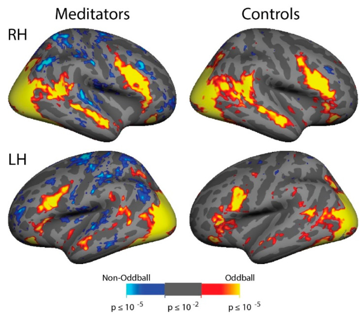
There were no significant differences between the experienced meditators and the matched controls in their performance of the attention tasks. But during the sustained attention task the meditators in comparison to the controls had increases in activation of the brain’s Dorsal Attentional Network at the same time as a significant decrease in the brain’s Default Mode Network. In addition, during the sustained attention task the meditators had significantly greater anticorrelations of activity between the two networks indicating higher levels of inhibitory activity between networks.


The Dorsal Attentional Network has been shown to be involved in focused attention. Hence, the results suggest that experience in meditation improves the response of the brain areas responsible for focused attention when needed. Alternatively, the Default Mode Network has been shown to be involved in mind wandering. Hence, the results suggest that experience in meditation reduces the response of the brain areas responsible a loss of focused attention and mind wandering when needed. Further, the results suggest that experience in meditation leads to a greater ability of the brain to focus attention while preventing mind wandering.


The fact that experienced meditators did not differ from controls in attentional task performance is a bit disappointing. But on average the meditators performed better but because of a small number of participants, the differences were not statistically significant. Perhaps with larger groups differences would be found. But the lack of statistical power makes the significant brain activation differences observed during sustained attention even more striking. This all suggests that meditation practice shapes the brain for better attentional ability.


So, meditation strengthens brain attentional network.


Meditation is an important form of self-control and a healthy practice. It augments focus and attention and could be used to enhance empathy and all attentional capacities.” – Gabriel José Corrêa Mograbi


CMCS – Center for Mindfulness and Contemplative Studies


This and other Contemplative Studies posts are also available on Google+ https://plus.google.com/106784388191201299496/posts and on Twitter @MindfulResearch


Study Summary


Devaney, K.J.; Levin, E.J.; Tripathi, V.; Higgins, J.P.; Lazar, S.W.; Somers, D.C. Attention and Default Mode Network Assessments of Meditation Experience during Active Cognition and Rest. Brain Sci. 2021, 11, 566. https://doi.org/10.3390/ brainsci11050566


Abstract: Meditation experience has previously been shown to improve performance on behavioral assessments of attention, but the neural bases of this improvement are unknown. Two prominent, strongly competing networks exist in the human cortex: a dorsal attention network, that is activated during focused attention, and a default mode network, that is suppressed during attentionally demanding tasks. Prior studies suggest that strong anti-correlations between these networks indicate good brain health. In addition, a third network, a ventral attention network, serves as a “circuit-breaker” that transiently disrupts and redirects focused attention to permit salient stimuli to capture attention. Here, we used functional magnetic resonance imaging to contrast cortical network activation between experienced focused attention Vipassana meditators and matched controls. Participants performed two attention tasks during scanning: a sustained attention task and an attention-capture task. Meditators demonstrated increased magnitude of differential activation in the dorsal attention vs. default mode network in a sustained attention task, relative to controls. In contrast, there were no evident attention network differences between meditators and controls in an attentional reorienting paradigm. A resting state functional connectivity analysis revealed a greater magnitude of anticorrelation between dorsal attention and default mode networks in the meditators as compared to both our local control group and a n = 168 Human Connectome Project dataset. These results demonstrate, with both task- and rest-based fMRI data, increased stability in sustained attention processes without an associated attentional capture cost in meditators. Task and resting-state results, which revealed stronger anticorrelations between dorsal attention and default mode networks in experienced mediators than in controls, are consistent with a brain health benefit of long-term meditation practice.

https://www.ncbi.nlm.nih.gov/pmc/articles/PMC8144977/


Improve Brain Processing in Mood Dysregulated Adolescents with Mindfulness

Improve Brain Processing in Mood Dysregulated Adolescents with Mindfulness


By John M. de Castro, Ph.D.


“Mindfulness-based interventions—practices that promote non-judgmental attention to the present—can help individuals respond with acceptance to challenging circumstances or emotions and is a promising approach to treatment of mood lability.” – D. M. Hafemann


Adolescence is a time of mental, physical, social, and emotional growth. But it can be a difficult time, fraught with challenges. During this time the child transitions to young adulthood; including the development of intellectual, psychological, physical, and social abilities and characteristics. There are so many changes occurring during this time that the child can feel overwhelmed and unable to cope with all that is required. This can lead to emotional and behavioral problems. Indeed, up to a quarter of adolescents suffer from depression or anxiety disorders, and an even larger proportion struggle with subclinical symptoms. Mindfulness training has been shown to improve emotion regulation and to benefit the psychological and emotional health of adolescents


The nervous system is a dynamic entity, constantly changing and adapting to the environment. It will change size, activity, and connectivity in response to experience. These changes in the brain are called neuroplasticity. Over the last decade neuroscience has been studying the effects of contemplative practices on the brain and has identified neuroplastic changes in widespread areas. In other words, mindfulness practice appears to mold and change the brain, producing psychological, physical, and spiritual benefits. The brains of adolescents are different from fully mature adult brains. They are dynamically growing and changing. It is unclear how mindfulness affects their maturing brains particularly in adolescents who have mood dysregulations.


In today’s Research News article “Network-level functional topological changes after mindfulness-based cognitive therapy in mood dysregulated adolescents at familial risk for bipolar disorder: a pilot study.” (See summary below or view the full text of the study at: https://www.ncbi.nlm.nih.gov/pmc/articles/PMC8080341/ ) Qin and colleagues recruited adolescents (13-17 years of age) who were mood dysregulated and who had at least one biological parent with bipolar disorder. They were provided a once a week for 75 minutes for 12-weeks program of Mindfulness-Based Cognitive Therapy (MBCT) adapted for children along with home practice. They were measured before and after training for emotion regulation, depressive and manic symptoms, overall global functioning, and clinical ratings. They also had their brains scanned with functional Magnetic Resonance Imaging (fMRI).


In comparison to baseline they found that after Mindfulness-Based Cognitive Therapy (MBCT) there were surprisingly no significant changes in emotion regulation, depressive and manic symptoms, overall global functioning, and clinical ratings. But there were significant increases in network efficiency and decreases in path length in the cingulo-opercular network and frontal parietal network and increases in the connectivity of brain structures within the cingulo-opercular network and the default mode network. In addition, the shorter the path length within the cingulo-opercular network the higher the level of emotion regulation.


These results need to be interpreted with caution as there was no control comparison condition and so there are potential confounding variables that could account for the results. But the psychological results are very disappointing. Mindfulness-Based Cognitive Therapy (MBCT) has been routinely found to improve emotions and emotion regulation in previous research. But it did not in the present study. It is possible that unlike with adults, MBCT is simply ineffective in improving the psychological health of mood dysregulated adolescents.


On the other hand, the functional Magnetic Resonance Imaging (fMRI) findings were interesting. The cingulo-opercular network and frontal parietal network are both involved in top-down cognitive control. The observed increases in network efficiency within these networks after Mindfulness-Based Cognitive Therapy (MBCT) suggests that MBCT improves the ability of mood dysregulated adolescents to control their thinking. This is exactly what MBCT is designed to do. Unfortunately, the researchers did not measure cognitive ability in this study, so there is no confirmatory behavioral results. The increased emotion regulation associated with decreases in path length in the cingulo-opercular network, though, suggests that the changes in the youths’ brains may be associated with improved ability to control their emotions. This may suggest an a lessened chance of developing major mental illness in the future.


So, Improve Brain Processing in Mood Dysregulated Adolescents with Mindfulness.


With low level of mindfulness, adolescents might be lack of emotion clarity, self-control and acceptance, which in turn might lead to their poor realization of emotion and easy immersion into dysfunctional emotional reactions such as impulse and aggressive behavior toward others or blame to themselves.” – Ying Ma


CMCS – Center for Mindfulness and Contemplative Studies


This and other Contemplative Studies posts are also available on Google+ https://plus.google.com/106784388191201299496/posts and on Twitter @MindfulResearch


Study Summary


Qin, K., Lei, D., Yang, J., Li, W., Tallman, M. J., Duran, L., Blom, T. J., Bruns, K. M., Cotton, S., Sweeney, J. A., Gong, Q., & DelBello, M. P. (2021). Network-level functional topological changes after mindfulness-based cognitive therapy in mood dysregulated adolescents at familial risk for bipolar disorder: a pilot study. BMC psychiatry, 21(1), 213. https://doi.org/10.1186/s12888-021-03211-4


Abstract

Background

Given that psychopharmacological approaches routinely used to treat mood-related problems may result in adverse outcomes in mood dysregulated adolescents at familial risk for bipolar disorder (BD), Mindfulness-Based Cognitive Therapy for Children (MBCT-C) provides an alternative effective and safe option. However, little is known about the brain mechanisms of beneficial outcomes from this intervention. Herein, we aimed to investigate the network-level neurofunctional effects of MBCT-C in mood dysregulated adolescents.

Methods

Ten mood dysregulated adolescents at familial risk for BD underwent a 12-week MBCT-C intervention. Resting-state functional magnetic resonance imaging (fMRI) was performed prior to and following MBCT-C. Topological metrics of three intrinsic functional networks (default mode network (DMN), fronto-parietal network (FPN) and cingulo-opercular network (CON)) were investigated respectively using graph theory analysis.

Results

Following MBCT-C, mood dysregulated adolescents showed increased global efficiency and decreased characteristic path length within both CON and FPN. Enhanced functional connectivity strength of frontal and limbic areas were identified within the DMN and CON. Moreover, change in characteristic path length within the CON was suggested to be significantly related to change in the Emotion Regulation Checklist score.

Conclusions

12-week MBCT-C treatment in mood dysregulated adolescents at familial risk for BD yield network-level neurofunctional effects within the FPN and CON, suggesting enhanced functional integration of the dual-network. Decreased characteristic path length of the CON may be associated with the improvement of emotion regulation following mindfulness training. However, current findings derived from small sample size should be interpreted with caution. Future randomized controlled trials including larger samples are critical to validate our findings.

https://www.ncbi.nlm.nih.gov/pmc/articles/PMC8080341/


Improve Nervous System and Cognitive Function with Tai Chi

Improve Nervous System and Cognitive Function with Tai Chi


By John M. de Castro, Ph.D.


“tai chi appears to improve executive function—the ability to multitask, manage time, and make decisions—in people without any cognitive decline. In those with mild cognitive impairment, tai chi slowed the progression to dementia.” – Harvard Health


Tai Chi is an ancient Chinese practice involving mindfulness and gentle movements. It is easy to learn, safe, and gentle. Tai Chi has been practiced for thousands of years with benefits for health and longevityTai Chi training is designed to enhance function and regulate the activities of the body through controlled breathing, mindful concentration, and gentle movements. Only recently though have the effects of this practice been scrutinized with empirical research. This research has found that it is effective for an array of physical and psychological issues. It appears to strengthen the immune systemreduce inflammation and increase the number of cancer killing cells in the bloodstream, improve cardiovascular healthreduce arthritis painimprove balance and reduce falls. It also appears to improve attentional ability improve cognitive ability in the elderly, and relieve depression.


The nervous system is a dynamic entity, constantly changing and adapting to the environment. It will change size, activity, and connectivity in response to experience. These changes in the brain are called neuroplasticity. Over the last decade neuroscience has been studying the effects of contemplative practices on the brain and has identified neuroplastic changes in widespread areas. In other words, mindfulness practice appears to mold and change the brain, producing psychological, physical, and spiritual benefits. Hence, it would appear likely that Tai Chi practice may alter the brain networks underlying mindfulness.


In today’s Research News article “Tai Chi Chuan Alters Brain Functional Network Plasticity and Promotes Cognitive Flexibility.” (See summary below or view the full text of the study at: https://www.frontiersin.org/articles/10.3389/fpsyg.2021.665419/full?utm_source=F-AAE&utm_medium=EMLF&utm_campaign=MRK_1671974_a0P58000000G0YfEAK_Psycho_20210701_arts_A ) Cui and colleagues recruited healthy adults and randomly assigned them to either a control condition or to 3, 60 minute sessions per week for 8 weeks of aerobic exercise (Brisk walking) or Tai Chi practice. Before and after practice they were measured for cognitive flexibility and they also had their brains scanned with functional Magnetic Resonance Imaging (fMRI).


They found that only after Tai Chi practice there was a significant increase in cognitive flexibility. The fMRI revealed that Tai Chi practice increased brain local efficiency compared with general aerobic exercise. Local efficiency reflects brain information transmission and processing in local densely interconnected areas. Tai Chi practice also increased the clustering coefficient of brain activity which reflects how well brain areas connect with other areas. Tai Chi practice increased Nodal global efficiency which reflects the efficiency of information transmission within brain networks. Importantly, they found that the higher the Nodal global efficiency the greater the level of cognitive flexibility. These changes with Tai Chi practice suggest that it increases brain specialization and that this is related to better cognitive ability.


A strength of the present study was that Tai Chi practice was compared to another aerobic exercise, brisk walking. So, it can be concluded that the effects observed were due to Tai Chi practice per se and not to the exercise provided by Tai Chi. The findings suggest then that practicing Tai Chi changes the brain making it better interconnected with a greater ability to share information and these changes are associated with greater cognitive flexibility. This shows that Tai Chi practice changes the brain in beneficial ways. This may be responsible for the ability of Tai Chi practice to improve the physical and psychological processes especially in the elderly.


So, improve nervous system and cognitive function with Tai Chi.


Tai Chi, a multicomponent mind-body exercise, combines slow physical activity with relaxation to serve as a movement meditation. Prior trials suggested that the beneficial effects of Tai Chi are created by a physical component which capitalizes on the benefits of physical exercise and a mind component which additionally promotes psychological well-being, life satisfaction, and improved perception of health.” – Chunlin Yue


CMCS – Center for Mindfulness and Contemplative Studies


This and other Contemplative Studies posts are also available on Google+ https://plus.google.com/106784388191201299496/posts and on Twitter @MindfulResearch


Study Summary


Cui L, Tao S, Yin H-c, Shen Q-q, Wang Y, Zhu L-n and Li X-j (2021) Tai Chi Chuan Alters Brain Functional Network Plasticity and Promotes Cognitive Flexibility. Front. Psychol. 12:665419. doi: 10.3389/fpsyg.2021.665419


Objective: This study used resting-state functional magnetic resonance imaging to investigate the effects of 8 weeks of Tai Chi Chuan and general aerobic exercise on the topological parameters of brain functional networks, explored the advantages of Tai Chi Chuan for improving functional network plasticity and cognitive flexibility, and examined how changes in topological attributes of brain functional networks relate to cognitive flexibility.

Methods: Thirty-six healthy adults were grouped into Tai Chi Chuan (Bafa Wubu of Tai Chi), general aerobic exercise (brisk walking), and control groups. All of the subjects underwent fMRI and behavioral assessment before and after the exercise intervention.

Results: Tai Chi Chuan exercise significantly enhanced the clustering coefficient and local efficiency compared with general aerobic exercise. Regarding the nodal properties, Tai Chi Chuan significantly enhanced the nodal clustering coefficient of the bilateral olfactory cortex and left thalamus, significantly reduced the nodal clustering coefficient of the left inferior temporal gyrus, significantly improved the nodal efficiency of the right precuneus and bilateral posterior cingulate gyrus, and significantly improved the nodal local efficiency of the left thalamus and right olfactory cortex. Furthermore, the behavioral performance results demonstrated that cognitive flexibility was enhanced by Tai Chi Chuan. The change in the nodal clustering coefficient in the left thalamus induced by Tai Chi Chuan was a significant predictor of cognitive flexibility.

Conclusion: These findings demonstrated that Tai Chi Chuan could promote brain functional specialization. Brain functional specialization enhanced by Tai Chi Chuan exercise was a predictor of greater cognitive flexibility.

https://www.frontiersin.org/articles/10.3389/fpsyg.2021.665419/full?utm_source=F-AAE&utm_medium=EMLF&utm_campaign=MRK_1671974_a0P58000000G0YfEAK_Psycho_20210701_arts_A


Meditation Increases Functional Connectivity of Brain Networks

Meditation Increases Functional Connectivity of Brain Networks


By John M. de Castro, Ph.D.


It seems the longer you do meditation, the better your brain will be at self-regulation,” – Bin He


Mindfulness training has been shown through extensive research to be effective in improving physical and psychological health and particularly with reducing the physical and psychological reactions to stress. There are a number of ways that meditation practices produce these benefits, including changes to the brain and physiology. The nervous system changes in response to how it is used and how it is stimulated in a process called neuroplasticity. Highly used areas grow in size, metabolism, and connectivity. Mindfulness practices in general are known to produce these kinds of changes in the structure and activity of the brain. There is little research, however, on how these changes develop with meditation.


In today’s Research News article “Longitudinal effects of meditation on brain resting-state functional connectivity.” (See summary below or view the full text of the study at: https://www.ncbi.nlm.nih.gov/pmc/articles/PMC8166909/ ) Zhang and colleagues recruited novice meditators enrolled in a university meditation course. They practiced focused attention meditation over 2 months twice a week in class and at home 5 times per week for 10 minutes. Before and after training the students had their brains scanned with functional Magnetic Resonance Imaging (f-MRI).


They found that after the 2-month meditation training the participants had significant increases in the functional connectivity within the Dorsal Attention Network of the brain and between the Dorsal Attention Network and the Default Mode Network and also between the Default Mode Network and the visual cortex.


The Dorsal Attention Network is a series of structures in the brain that are associated with attentional focusing while the Default Mode Network is a series of structures in the brain that are associated with self-referential thought and mind wandering. Typically, during focused attention meditation, particularly in novice meditators, the mind switches back and forth between focus on the object of meditation and unfocused mind wandering. During mind wandering, visualizations of this content often occur. This usually occurs repeatedly during the meditation session. This switching involves going back and forth between the Dorsal Attention Network and the Default Mode Network and the visual areas of the brain. The brain scan findings indicate that this results in an increase in the functional connectivity between the two networks. Hence, the changes in the mental contents during meditation are reflected in brain activity.


So, meditation increases functional connectivity of brain networks.


So, not only did meditation change the structures in the participants’ brains, it also changed how they felt.” – Lela Moore


CMCS – Center for Mindfulness and Contemplative Studies


This and other Contemplative Studies posts are also available on Google+ https://plus.google.com/106784388191201299496/posts and on Twitter @MindfulResearch


Study Summary


Zhang, Z., Luh, W. M., Duan, W., Zhou, G. D., Weinschenk, G., Anderson, A. K., & Dai, W. (2021). Longitudinal effects of meditation on brain resting-state functional connectivity. Scientific reports, 11(1), 11361. https://doi.org/10.1038/s41598-021-90729-y


Abstract

Changes in brain resting-state functional connectivity (rsFC) were investigated using a longitudinal design by following a 2-month focused attention meditation (FAM) practice and analyzing their association with FAM practice time. Ten novice meditators were recruited from a university meditation course. Participants were scanned with a resting-state fMRI sequence with multi-echo EPI acquisition at baseline and at the 2-month follow-up. Total FAM practice time was calculated from the daily log of the participants. We observed significantly increased rsFC between the posterior cingulate cortex (PCC) and dorsal attention network (DAN), the right middle temporal (RMT) region and default mode network (DMN), the left and right superior parietal lobules (LSPL/RSPL) and DMN, and the LSPL/RSPL and DAN. Furthermore, the rsFC between the LSPL and medial prefrontal cortex was significantly associated with the FAM practice time. These results demonstrate increased connectivity within the DAN, between the DMN and DAN, and between the DMN and visual cortex. These findings demonstrate that FAM can enhance the brain connection among and within brain networks, especially DMN and DAN, indicating potential effect of FAM on fast switching between mind wandering and focused attention and maintaining attention once in the attentive state.

https://www.ncbi.nlm.nih.gov/pmc/articles/PMC8166909/


Different Meditation Styles Affect the Medial Frontal Brain Network Differently

Different Meditation Styles Affect the Medial Frontal Brain Network Differently


By John M. de Castro, Ph.D.


“meditation has a variety of neurological benefits, from changes in brain volume to decreasing activity in parts of the brain involved with stress.” – Ashley Welch


Mindfulness training has been shown to improve health and well-being. It has also been found to be effective for a large array of medical and psychiatric conditions, either stand-alone or in combination with more traditional therapies. How exactly mindfulness practices produce their benefits is unknown. But it is known that meditation practice alters brain activity.


There are a number of different types of meditation. Classically they’ve been characterized on a continuum with the degree and type of attentional focus. In focused attention meditation, the individual practices paying attention to a single meditation object, usually the breath. In open monitoring meditation, the individual opens up awareness to everything that’s being experienced including thoughts regardless of its origin. In Loving Kindness Meditation the individual

systematically pictures different individuals from self, to close friends, to enemies and wishes them happiness, well-being, safety, peace, and ease of well-being. It is suspected but not known that different forms of meditation practice can produce different changes in brain activity.


One way is to measure changes in the electroencephalogram (EEG), the rhythmic electrical activity that can be recorded from the scalp. In today’s Research News article “Attentional and cognitive monitoring brain networks in long-term meditators depend on meditation states and expertise.” (See summary below or view the full text of the study at: https://www.ncbi.nlm.nih.gov/pmc/articles/PMC7921394/ )  Yordanova and colleagues recruited highly experienced meditators who practiced focused attention meditation, open monitoring meditation, and Loving Kindness Meditation in a balanced way. They had their electroencephalograms (EEG) recorded while at rest and while performing the 3 meditation types for 3 minutes each.


They found that the Frontal-Parietal network, that is thought to underlie attentional mechanisms did not differ between meditation types. But there was increased connectivity between the right hemisphere frontal and left hemisphere parietal areas. On the other hand, the Medial Frontal network that is thought to underlie cognitive control and monitoring mechanisms had different activity patterns with the different meditation types. During focused attention meditation was increased synchronization in the parietal regions whereas during Loving Kindness Meditation it increased in the right frontal regions.


These are interesting findings that demonstrate that highly experienced meditators have distinct changes in the activity of their brains during meditation regardless of type. But in areas associated with cognitive monitoring mechanisms, difference appear. During focused attention meditation and Loving Kindness Meditation there are different patterns of activity. To some extent this is not surprising in that the two meditation types involve specific focuses. But Loving Kindness Meditation is emotionally focused while focused attention meditation is breath sensation focused and these require different kinds of cognitive control. These differences may underlie the different medial frontal activities.


It should be noted that these patterns are quite different from those of inexperienced meditators and that the greater the amount of practice the greater the neural activations. It would be expected that highly experienced meditators would have greater focus and much less mind wandering during meditation than inexperience meditators and this would produce different patterns of neural activation.


So, different meditation styles affect the medial frontal brain network differently.


Meditation benefits for the brain are abundant. Meditating strengthens neural connections and can literally change the configuration of these networks. With regular practice, you can cultivate a more resilient neurobiology.” – Ask the Scientists


CMCS – Center for Mindfulness and Contemplative Studies


This and other Contemplative Studies posts are also available on Google+ https://plus.google.com/106784388191201299496/posts and on Twitter @MindfulResearch


Study Summary


Yordanova, J., Kolev, V., Nicolardi, V., Simione, L., Mauro, F., Garberi, P., Raffone, A., & Malinowski, P. (2021). Attentional and cognitive monitoring brain networks in long-term meditators depend on meditation states and expertise. Scientific reports, 11(1), 4909. https://doi.org/10.1038/s41598-021-84325-3


Abstract

Meditation practice is suggested to engage training of cognitive control systems in the brain. To evaluate the functional involvement of attentional and cognitive monitoring processes during meditation, the present study analysed the electroencephalographic synchronization of fronto-parietal (FP) and medial-frontal (MF) brain networks in highly experienced meditators during different meditation states (focused attention, open monitoring and loving kindness meditation). The aim was to assess whether and how the connectivity patterns of FP and MF networks are modulated by meditation style and expertise. Compared to novice meditators, (1) highly experienced meditators exhibited a strong theta synchronization of both FP and MF networks in left parietal regions in all mediation styles, and (2) only the connectivity of lateralized beta MF networks differentiated meditation styles. The connectivity of intra-hemispheric theta FP networks depended non-linearly on meditation expertise, with opposite expertise-dependent patterns found in the left and the right hemisphere. In contrast, inter-hemispheric FP connectivity in faster frequency bands (fast alpha and beta) increased linearly as a function of expertise. The results confirm that executive control systems play a major role in maintaining states of meditation. The distinctive lateralized involvement of FP and MF networks appears to represent a major functional mechanism that supports both generic and style-specific meditation states. The observed expertise-dependent effects suggest that functional plasticity within executive control networks may underpin the emergence of unique meditation states in expert meditators.

https://www.ncbi.nlm.nih.gov/pmc/articles/PMC7921394/


Meditation Changes the Brain Areas Underlying Executive Function and Mind Wandering

Meditation Changes the Brain Areas Underlying Executive Function and Mind Wandering


By John M. de Castro, Ph.D.


“meditation nurtures the parts of the brain that contribute to well-being. Furthermore, it seems that a regular practice deprives the stress and anxiety-related parts of the brain of their nourishment.” – Mindworks


Mindfulness training has been shown to improve health and well-being. It has also been found to be effective for a large array of medical and psychiatric conditions, either stand-alone or in combination with more traditional therapies. There are a number of ways that mindfulness practices produce these benefits, including changes to the brain and physiology. The nervous system is a dynamic entity, constantly changing and adapting to the environment. It will change size, activity, and connectivity in response to experience. These changes in the brain are called neuroplasticity. Over the last decade neuroscience has been studying the effects of contemplative practices on the brain and has identified neuroplastic changes in widespread areas. In other words, mindfulness practice appears to mold and change the brain, producing psychological, physical, and spiritual benefits.


In today’s Research News article “Resting State Functional Connectivity Associated With Sahaja Yoga Meditation.” (See summary below or view the full text of the study at: https://www.ncbi.nlm.nih.gov/pmc/articles/PMC8007769/ )  Barrós-Loscertales and colleagues recruited healthy adult non-meditators and long-term practitioners of Sahaja Yoga Meditation. They were measured for impulsiveness and cognitive control and response inhibition with a go/no-go task in which the participants had to respond to a target stimulus and withhold responding to another stimulus and a Simon task in which they had to respond to a target stimulus pointing in the same direction as its location and withhold responding to another stimulus pointing in a different direction than its location. The participants also had their brains scanned with functional Magnetic Resonance Imaging (fMRI).


They found that the meditators had increased functional connectivity between the ventrolateral prefrontal cortex and the dorsolateral prefrontal cortex and decreased functional connectivity between the left insula and mid-cingulate cortex and between the right angular gyrus and precuneus. They also found that the meditators had significantly less response interference and the greater the amount of response interference the lower the functional connectivity between the left insula and the mid-cingulate.


The ventrolateral prefrontal cortex and the dorsolateral prefrontal cortex have been found to be involved in attention, cognitive control, and conflict resolution. The increased connectivity suggests that the meditators had higher levels of executive function. The insula, cingulate cortex, angular gyrus, and precuneus are all components of the default mode network that has been shown to be involved with mind wandering and self-referential thinking. The decreased functional connectivity between these areas suggests reductions in these processes.


It should be kept in mind that the meditation and non-meditation groups self-selected whether they meditated or not and thus may be systematically different people. So, causation cannot be determined. Nevertheless, these results suggest that the meditators had more active attention and executive control networks and less active mind-wandering networks. These results replicate previous research that demonstrated that contemplative practices like meditation produce improvements in cognition and reductions in mind wandering.  So, the connectivity differences seen in the present study appear to show the neural mechanisms underlying the differences in thought processes.


So, meditation changes the brain areas underlying executive function and mind wandering.


It seems the longer you do meditation, the better your brain will be at self-regulation. You don’t have to consume as much energy at rest and you can more easily get yourself into a more relaxed state.” – Bin He


CMCS – Center for Mindfulness and Contemplative Studies


This and other Contemplative Studies posts are also available on Google+ https://plus.google.com/106784388191201299496/posts and on Twitter @MindfulResearch


Study Summary


Barrós-Loscertales, A., Hernández, S. E., Xiao, Y., González-Mora, J. L., & Rubia, K. (2021). Resting State Functional Connectivity Associated With Sahaja Yoga Meditation. Frontiers in human neuroscience, 15, 614882. https://doi.org/10.3389/fnhum.2021.614882


Abstract

Neuroscience research has shown that meditation practices have effects on brain structure and function. However, few studies have combined information on the effects on structure and function in the same sample. Long-term daily meditation practice produces repeated activity of specific brain networks over years of practice, which may induce lasting structural and functional connectivity (FC) changes within relevant circuits. The aim of our study was therefore to identify differences in FC during the resting state between 23 Sahaja Yoga Meditation experts and 23 healthy participants without meditation experience. Seed-based FC analysis was performed departing from voxels that had shown structural differences between these same participants. The contrast of connectivity maps yielded that meditators showed increased FC between the left ventrolateral prefrontal cortex and the right dorsolateral prefrontal cortex but reduced FC between the left insula and the bilateral mid-cingulate as well as between the right angular gyrus and the bilateral precuneus/cuneus cortices. It thus appears that long-term meditation practice increases direct FC between ventral and dorsal frontal regions within brain networks related to attention and cognitive control and decreases FC between regions of these networks and areas of the default mode network.

https://www.ncbi.nlm.nih.gov/pmc/articles/PMC8007769/


Increase the Energy Metabolism of the Brain with Meditation

Increase the Energy Metabolism of the Brain with Meditation


By John M. de Castro, Ph.D.


As a form of mental training, meditation improves core physical and psychological assets, including energy, motivation, and strength. Studies on the neurophysiological concomitants of meditation have proved that commitment to daily practice can bring promising changes for the mind and the body.” –  Madhuleena Roy Chowdhury


Mindfulness training has been shown through extensive research to be effective in improving physical and psychological health and particularly with reducing the physical and psychological reactions to stress. There are a number of ways that meditation practices produce these benefits, including changes to the brain and physiology. The nervous system changes in response to how it is used and how it is stimulated in a process called neuroplasticity. Highly used areas grow in size, metabolism, and connectivity. Mindfulness practices in general are known to produce these kinds of changes in the structure and activity of the brain.


In today’s Research News article “Short-term meditation training influences brain energy metabolism: A pilot study on 31 P MR spectroscopy.” (See summary below or view the full text of the study at: https://www.ncbi.nlm.nih.gov/pmc/articles/PMC7821578/ ) Gizewski and colleagues recruited healthy adult meditation naïve yoga students and provided them with 7 weeks of twice a week 45-minute training in Raja yoga meditation. This focused meditation training emphasizes the cessation of thinking and includes meditation and breathing exercises. They were measured before and after training for meditative depth, health history, lifestyle, anxiety, depression, and angst. Before and after training they also underwent brain scanning with structural Magnetic Resonance Imaging (MRI) and for brain energy metabolism (31P-MRS).


They found that in comparison to baseline after Raja yoga meditation training there were significant increases overall mental health and decreases in anxiety and dysthymia. There were also significant increases in brain energy metabolism particularly in the right hemisphere in the occipital and temporal lobes and the basal ganglia.


This study did not contain a comparison, control, condition which opens the results up to some alternative interpretations. But ignoring these possible contaminants, the study suggests that 7 weeks of meditation training can alter the brain. This has been demonstrated with numerous studies of changes in the structure, connectivity, and electrical activity of the brain produced by mindfulness training. The present study adds to this understanding by demonstrating the focused meditation training increases the energy metabolism in the brain particularly in the posterior cerebral cortex and the motor control areas. Meditation training is thought to be relaxing and the technique used here is one that emphasizes reduction in mental activity. But the present study suggests that the brain can get very active. This suggests that there is considerable mental activity going on during meditation.


So, increase the energy metabolism of the brain with meditation.


Meditation is thought to work via its effects on the sympathetic nervous system, which increases heart rate, breathing, and blood pressure during times of stress. Yet meditating has a spiritual purpose, too. “True, it will help you lower your blood pressure, but so much more: it can help your creativity, your intuition, your connection with your inner self,” –  Burke Lennihan,


CMCS – Center for Mindfulness and Contemplative Studies


This and other Contemplative Studies posts are also available on Google+ https://plus.google.com/106784388191201299496/posts and on Twitter @MindfulResearch


Study Summary


Gizewski, E. R., Steiger, R., Waibel, M., Pereverzyev, S., Sommer, P., Siedentopf, C., Grams, A. E., Lenhart, L., & Singewald, N. (2021). Short-term meditation training influences brain energy metabolism: A pilot study on 31 P MR spectroscopy. Brain and behavior, 11(1), e01914. https://doi.org/10.1002/brb3.1914


Abstract

Background

Meditation is increasingly attracting interest among neuroimaging researchers for its relevance as a cognitive enhancement technique and several cross‐sectional studies have indicated cerebral changes. This longitudinal study applied a distinct and standardized meditative technique with a group of volunteers in a short‐term training program to analyze brain metabolic changes.

Methods

The effect of 7 weeks of meditation exercises (focused attention meditation, FAM) was assessed on 27 healthy volunteers. Changes in cerebral energy metabolism were investigated using 31P‐MR spectroscopy. Metabolite ratios were compared before (T1) and after training (T2). Additional questionnaire assessments were included.

Results

The participants performed FAM daily. Depression and anxiety scores revealed a lower level of state anxiety at T2 compared to T1. From T1 to T2, energy metabolism ratios showed the following differences: PCr/ATP increased right occipitally; Pi/ATP decreased bilaterally in the basal ganglia and temporal lobe on the right; PCr/Pi increased in occipital lobe bilaterally, in the basal ganglia and in the temporal lobe on the right side. The pH decreased temporal on the left side and frontal in the right side. The observed changes in the temporal areas and basal ganglia may be interpreted as a higher energetic state, whereas the frontal and occipital areas showed changes that may be related to a down‐regulation in ATP turnover, energy state, and oxidative capacity.

Conclusions

The results of the current study indicate for the first time in a longitudinal study that even short‐term training in FAM may have considerable effects on brain energy state with different local energy management in specific brain regions. Especially higher energetic state in basal ganglia may represent altered function in their central role in complex cerebral distributed networks including frontal and temporal areas. Further studies including different forms of relaxation techniques should be performed for more specific and reliable insights.

https://www.ncbi.nlm.nih.gov/pmc/articles/PMC7821578/


Improve Brain Function and Mental Health in Chronic Pain Patients with Mindfulness

Improve Brain Function and Mental Health in Chronic Pain Patients with Mindfulness


By John M. de Castro, Ph.D.


“With the ACT approach, learning to let go of behaviors, memories, thoughts, and emotions creates space to explore new experiences, including acceptance of chronic pain sensations.” – Rosemary Black

We all have to deal with pain. It’s inevitable, but hopefully it’s mild and short lived. For a wide swath of humanity, however, pain is a constant in their lives. At least 100 million adult Americans have chronic pain conditions. The most common treatment for chronic pain is drugs. These include over-the-counter analgesics and opioids. But opioids are dangerous and highly addictive. Prescription opioid overdoses kill more than 14,000 people annually. So, there is a great need to find safe and effective ways to lower the psychological distress and improve the individual’s ability to cope with the pain.


Hence, it is important to find an effective method to treat Chronic pain. There is an accumulating volume of research findings to demonstrate that mindfulness practices, in general, are effective in treating pain. Acceptance and Commitment Therapy (ACT) is a mindfulness-based psychotherapy technique that is employs many of the techniques of Cognitive Behavioral Therapy (CBT). ACT focuses on the individual’s thoughts, feelings, and behavior and how they interact to impact their psychological and physical well-being. It then works to change thinking to alter the interaction and produce greater life satisfaction. ACT employs mindfulness practices to increase awareness and develop an attitude of acceptance and compassion in the presence of painful thoughts and feelings. ACT teaches individuals to “just notice”, accept and embrace private experiences and focus on behavioral responses that produce more desirable outcomes.


Pain experiences are processed in the nervous system. So, it’s likely that mindfulness practices somehow alter the brain’s processing of pain. So, it makes sense to study the how Acceptance and Commitment Therapy (ACT) may affect the brain to improve the mental health of chronic pain patients.


In today’s Research News article “Neural Mechanisms of Acceptance and Commitment Therapy for Chronic Pain: A Network-Based fMRI Approach.” (See summary below or view the full text of the study at: https://www.ncbi.nlm.nih.gov/pmc/articles/PMC7892587/ ) Aytur and colleagues recruited with musculoskeletal pain who did not abuse opioids. They completed 2 sessions per week for 4 weeks of Acceptance and Commitment Therapy (ACT). Before and after therapy they were measured for acceptance, chronic pain acceptance, quality of life, pain interference, and depression. Brain systems were measured before and after therapy with functional Magnetic Resonance Imaging (fMRI).


They found that in comparison to baseline after Acceptance and Commitment Therapy (ACT) the participants had significant increases in acceptance, chronic pain acceptance, quality of life and significant decreases in pain interference, and depression. In the fMRIs they also observed significant decreases in activation and connectivity in the Default Mode Network (DMN), Salience Network (SN), and Frontal-Parietal Network (FPN) networks of the brain. In addition, they observed that changes in the connectivity between these networks were correlated with decreases in depression and pain interference, and increases in quality of life.


These are interesting results that need to be interpreted with caution as there wasn’t a control, comparison condition present opening the findings up to alternative confounded interpretations. But the results replicate previous findings with controlled designs that Acceptance and Commitment Therapy (ACT) produces improvements in depression, pain, and quality of life. So, the improvements observed in the present study are likely due to causal effects of ACT.


These results also show that the psychological improvements produced by Acceptance and Commitment Therapy (ACT) are related to changes in the activity and connectivity within and between significant brain networks. This suggests that these neural alterations may underly the improvements in mental health of chronic pain patients produced by ACT. It remains for future studies to unravel the cause effect relationships.


So, improve brain function and mental health in chronic pain patients with mindfulness.


ACT can help us to break out of that box that we have created for ourselves. To understand that the key to living with our condition is not to limit our lives and miss out on experiences, but instead to face things head on and be dedicated to working with our pain and doing things that bring us joy. ACT’s goal is not necessarily to reduce the pain but to help you to live your life with it, to feel more confident in engaging in activities that you might have otherwise swayed away from.” – Ann-Marie D’arcy-Sharpe


CMCS – Center for Mindfulness and Contemplative Studies


This and other Contemplative Studies posts are also available on Google+ https://plus.google.com/106784388191201299496/posts and on Twitter @MindfulResearch


Study Summary


Aytur, S. A., Ray, K. L., Meier, S. K., Campbell, J., Gendron, B., Waller, N., & Robin, D. A. (2021). Neural Mechanisms of Acceptance and Commitment Therapy for Chronic Pain: A Network-Based fMRI Approach. Frontiers in human neuroscience, 15, 587018. https://doi.org/10.3389/fnhum.2021.587018


Abstract

Over 100 million Americans suffer from chronic pain (CP), which causes more disability than any other medical condition in the United States at a cost of $560–$635 billion per year (Institute of Medicine, 2011). Opioid analgesics are frequently used to treat CP. However, long term use of opioids can cause brain changes such as opioid-induced hyperalgesia that, over time, increase pain sensation. Also, opioids fail to treat complex psychological factors that worsen pain-related disability, including beliefs about and emotional responses to pain. Cognitive behavioral therapy (CBT) can be efficacious for CP. However, CBT generally does not focus on important factors needed for long-term functional improvement, including attainment of personal goals and the psychological flexibility to choose responses to pain. Acceptance and Commitment Therapy (ACT) has been recognized as an effective, non-pharmacologic treatment for a variety of CP conditions (Gutierrez et al., 2004). However, little is known about the neurologic mechanisms underlying ACT. We conducted an ACT intervention in women (n = 9) with chronic musculoskeletal pain. Functional magnetic resonance imaging (fMRI) data were collected pre- and post-ACT, and changes in functional connectivity (FC) were measured using Network-Based Statistics (NBS). Behavioral outcomes were measured using validated assessments such as the Acceptance and Action Questionnaire (AAQ-II), the Chronic Pain Acceptance Questionnaire (CPAQ), the Center for Epidemiologic Studies Depression Scale (CES-D), and the NIH Toolbox Neuro-QoLTM (Quality of Life in Neurological Disorders) scales. Results suggest that, following the 4-week ACT intervention, participants exhibited reductions in brain activation within and between key networks including self-reflection (default mode, DMN), emotion (salience, SN), and cognitive control (frontal parietal, FPN). These changes in connectivity strength were correlated with changes in behavioral outcomes including decreased depression and pain interference, and increased participation in social roles. This study is one of the first to demonstrate that improved function across the DMN, SN, and FPN may drive the positive outcomes associated with ACT. This study contributes to the emerging evidence supporting the use of neurophysiological indices to characterize treatment effects of alternative and complementary mind-body therapies.

https://www.ncbi.nlm.nih.gov/pmc/articles/PMC7892587/


Maintain Weight Loss with Mindfulness

Maintain Weight Loss with Mindfulness


By John M. de Castro, Ph.D.


“Just as meditation can help us with stress, sleeping, focus, and much more, it can also have an impact on our relationship with eating and managing our weight.” – Headspace


Eating is produced by two categories of signals. Homeostatic signals emerge from the body’s need for nutrients, is associated with feelings of hunger, and usually work to balance intake with expenditure. Non-homeostatic eating, on the other hand, is not tied to nutrient needs or hunger but rather to the environment, to emotional states, and or to the pleasurable and rewarding qualities of food. These cues can be powerful signals to eat even when there is no physical need for food. External eating is non-homeostatic eating in response to the environmental stimuli that surround us, including the sight and smell of food or the sight of food related cause such as the time of day or a fast-food restaurant ad or sign.


Mindful eating involves paying attention to eating while it is occurring, including attention to the sight, smell, flavors, and textures of food, to the process of chewing and may help reduce intake. Indeed, high levels of mindfulness are associated with lower levels of obesity and mindfulness training has been shown to reduce binge eating, emotional eating, and external eating. The Mindfulness-Based Stress Reduction (MBSR) program is a mindfulness program including meditation, body scan, yoga, discussion and home practice. So, it makes sense to examine MBSR as a part of a weight loss program.


In today’s Research News article “Keeping weight off: Mindfulness-Based Stress Reduction alters amygdala functional connectivity during weight loss maintenance in a randomized control trial.” (See summary below or view the full text of the study at: https://www.ncbi.nlm.nih.gov/pmc/articles/PMC7799782/ ) Chumachenko and colleagues recruited overweight adults (BMI>25) who had lost at least 5% of their body weight and wished to maintain it off. They were randomly assigned to receive 8, once a week for 1.5 hours, sessions of either a healthy living course or the Mindfulness-Based Stress Reduction (MBSR) program. They were measured before and after training and 6 months later for body size, perceived stress, depression, and eating disinhibition. Before and after training they also had their brains scanned with functional Magnetic Resonance Imaging (fMRI).


They found that after Mindfulness-Based Stress Reduction (MBSR) training there was a significant increase in the connectivity between the Amygdala and the ventromedial prefrontal cortex while this was decreased after completing the healthy living course. Over the 6-month follow-up period the MBSR participants did not gain weight while the healthy living course participants gained on average 5.9 pounds.


These results are interesting and important. There are many dietary programs that produce weight loss. But almost inevitably the weight is regained subsequently. Mindfulness-Based Stress Reduction (MBSR) training was found here to prevent that weight gain. Hence, MBSR may be an important ingredient in weight loss to help maintain the loss. The results also suggest that MBSR may alter the brain, increasing the functional connectivity in brain circuits that are thought to underlie emotion regulation. This fits with the prior findings that mindfulness training improves emotion regulation. This suggests that MBSR may prevent non-homeostatic, emotional, eating by strengthening emotion regulation and thereby prevent weight regain.


So, maintain weight loss with mindfulness.


By itself, mindful eating is not a weight-loss cure, but as part of an approach or tool it can catapult healthy eating and weight loss,” – Josh Klapow


CMCS – Center for Mindfulness and Contemplative Studies


This and other Contemplative Studies posts are also available on Google+ https://plus.google.com/106784388191201299496/posts and on Twitter @MindfulResearch


Study Summary


Chumachenko, S. Y., Cali, R. J., Rosal, M. C., Allison, J. J., Person, S. J., Ziedonis, D., Nephew, B. C., Moore, C. M., Zhang, N., King, J. A., & Fulwiler, C. (2021). Keeping weight off: Mindfulness-Based Stress Reduction alters amygdala functional connectivity during weight loss maintenance in a randomized control trial. PloS one, 16(1), e0244847. https://doi.org/10.1371/journal.pone.0244847


Abstract

Obesity is associated with significant comorbidities and financial costs. While behavioral interventions produce clinically meaningful weight loss, weight loss maintenance is challenging. The objective was to improve understanding of the neural and psychological mechanisms modified by mindfulness that may predict clinical outcomes. Individuals who intentionally recently lost weight were randomized to Mindfulness-Based Stress Reduction (MBSR) or a control healthy living course. Anthropometric and psychological factors were measured at baseline, 8 weeks and 6 months. Functional connectivity (FC) analysis was performed at baseline and 8 weeks to examine FC changes between regions of interest selected a priori, and independent components identified by independent component analysis. The association of pre-post FC changes with 6-month weight and psychometric outcomes was then analyzed. Significant group x time interaction was found for FC between the amygdala and ventromedial prefrontal cortex, such that FC increased in the MBSR group and decreased in controls. Non-significant changes in weight were observed at 6 months, where the mindfulness group maintained their weight while the controls showed a weight increase of 3.4% in BMI. Change in FC at 8-weeks between ventromedial prefrontal cortex and several ROIs was associated with change in depression symptoms but not weight at 6 months. This pilot study provides preliminary evidence of neural mechanisms that may be involved in MBSR’s impact on weight loss maintenance that may be useful for designing future clinical trials and mechanistic studies.

https://www.ncbi.nlm.nih.gov/pmc/articles/PMC7799782/


The Default Mode Network of the Brain Underlies Mind Wandering

The Default Mode Network of the Brain Underlies Mind Wandering


By John M. de Castro, Ph.D.


“the brain appears to support mind wandering by disrupting some of the brain processes that are involved in responding to our surrounding external environment.” – Julia Kam


We spend a tremendous amount of waking time with our minds wandering and not on the present environment or the task at hand. We daydream, plan for the future, review the past, ruminate on our failures, exalt in our successes. In fact, we spend almost half of our waking hours off task with our mind wandering. A system of the brain known as the Default Mode Network (DMN) becomes active during wind wandering and relatively quiet during focused on task behavior. Meditation is known to reduce the size, connectivity, and activity of the Default Mode Network (DMN).


In today’s Research News article “Lesion network mapping demonstrates that mind-wandering is associated with the default mode network.” (See summary below or view the full text of the study at: https://www.ncbi.nlm.nih.gov/pmc/articles/PMC7704688/ ) Philippi and colleagues recruited patients with circumscribed brain injuries (lesions) and age and education matched non-brain damaged comparison participants. They all underwent brain scanning with Magnetic Resonance Imaging (MRI). They completed a self-report measure of mind wandering.


They found that the brain damaged patients had lower frequencies of mind wandering than the healthy comparison participants. They then examined the specific brain areas damaged in the patients lesions and related it to their reduced mind wandering scores. They found that reduced mind wandering was associated with structures in the Default Mode Network (DMN), including the medial prefrontal cortex, parietal lobe, and inferior frontal gyrus.


The results are simple and straightforward and suggest that damage to the Default Mode Network (DMN) reduces mind wandering. This finding taken together with the findings that the DMN becomes more active during mind wandering makes a clear case that the DMN is responsible for mind wandering.


So, the default mode network of the brain underlies mind wandering.


mind-wandering was associated with increased DMN activity and increased DMN-VS connectivity.“ – Xinqi Zhou


CMCS – Center for Mindfulness and Contemplative Studies


This and other Contemplative Studies posts are also available on Google+ https://plus.google.com/106784388191201299496/posts and on Twitter @MindfulResearch


Study Summary


Philippi, C. L., Bruss, J., Boes, A. D., Albazron, F. M., Deifelt Streese, C., Ciaramelli, E., Rudrauf, D., & Tranel, D. (2021). Lesion network mapping demonstrates that mind-wandering is associated with the default mode network. Journal of neuroscience research, 99(1), 361–373. https://doi.org/10.1002/jnr.24648


Abstract

Functional neuroimaging research has consistently associated brain structures within the default mode network (DMN) and frontoparietal network (FPN) with mind-wandering. Targeted lesion research has documented impairments in mind-wandering after damage to the medial prefrontal cortex (mPFC) and hippocampal regions associated with the DMN. However, no lesion studies to date have applied lesion network mapping to identify common networks associated with deficits in mind-wandering. In lesion network mapping, resting-state functional connectivity data from healthy participants are used to infer which brain regions are functionally connected to each lesion location from a sample with brain injury. In the current study, we conducted a lesion network mapping analysis to test the hypothesis that lesions affecting the DMN and FPN would be associated with diminished mind-wandering. We assessed mind-wandering frequency on the Imaginal Processes Inventory (IPI) in participants with brain injury (n = 29) and healthy comparison participants without brain injury (n = 19). Lesion network mapping analyses showed the strongest association of reduced mind-wandering with the left inferior parietal lobule within the DMN. In addition, traditional lesion symptom mapping results revealed that reduced mind-wandering was associated with lesions of the dorsal, ventral, and anterior sectors of mPFC, parietal lobule, and inferior frontal gyrus in the DMN (p < 0.05 uncorrected). These findings provide novel lesion support for the role of the DMN in mind-wandering and contribute to a burgeoning literature on the neural correlates of spontaneous cognition.

Significance

Adults spend up to 50% of their waking day mind-wandering, which is the process of turning one’s attention inward to focus on self-generated thoughts or feelings. Mind-wandering can have both costs and benefits, such as increased negative mood or enhanced creative problem-solving. In this study, we report novel findings linking reduced mind-wandering with brain injury located within the default mode network. This work is important because it can help us to determine which brain networks are necessary for self-generated cognition, which may improve our understanding of neuropsychiatric conditions associated with altered self-focused thought.

https://www.ncbi.nlm.nih.gov/pmc/articles/PMC7704688/