Spirituality May Alter the Brain to Protect Against Major Depression

Spirituality May Alter the Brain to Protect Against Major Depression


By John M. de Castro, Ph.D.


“spirituality or religion may protect against major depression by thickening the brain cortex and counteracting the cortical thinning that would normally occur with major depression.” – Lisa Miller


Spirituality is defined as “one’s personal affirmation of and relationship to a higher power or to the sacred. Spirituality has been promulgated as a solution to the challenges of life both in a transcendent sense and in a practical sense. There have been a number of studies of the influence of spirituality on the physical and psychological well-being of practitioners mostly showing positive benefits, with spirituality encouraging personal growth and mental health.


One way that spirituality can have its effects on the individual is by altering the brain. The nervous system is a dynamic entity, constantly changing and adapting to the environment. It will change size, activity, and connectivity in response to experience. These changes in the brain are called neuroplasticity.  Over the last decade neuroscience has been studying the effects of contemplative practices on the brain and has identified neuroplastic changes in widespread area. and have found that meditation practice appears to mold and change the brain, producing psychological, physical, and spiritual benefits. So, religion and spirituality may be associated with changes in the nervous system associated with better mental health.


In today’s Research News article “Altruism and “love of neighbor” offer neuroanatomical protection against depression. Psychiatry research.” (See summary below or view the full text of the study at: https://www.ncbi.nlm.nih.gov/pmc/articles/PMC8672211/ ) Miller and colleagues reanalyzed longitudinal data obtained from individuals at risk for major depression and matched normal participants. At 30 and 35 years of age the participants brains were scanned with Magnetic Resonance Imaging (MRI) and the participants completed measures of major depressive disorder, level of depression and spirituality including measures of altruism, love thy neighbor as self, interconnectedness, contemplative practice, and commitment to religion/spirituality.


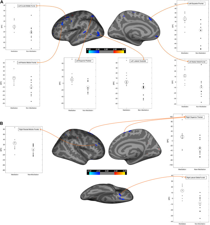
They found that the low risk of depression group had significantly greater cortical thickness in the Ventral Frontotemporal Network (VFTN), in comparison to the high-risk group. The VFTN had been previously shown to be associated with spiritual experience. They also found that in the at-risk for major depression group the greater the cortical thickness in the VFTN the lower levels of depression and the lower the risk of developing major depressive disorder. Across all participants, the higher the spirituality measures of altruism and love thy neighbor as self the greater the cortical thickness in the VFTN. In addition, in the high-risk group, the higher the levels of the spirituality measure of love thy neighbor as self the lower the levels of depression and the lower the risk of developing major depressive disorder.


The results demonstrate that the thickness of the Ventral Frontotemporal Network (VFTN) is associated with lower levels of depression and risk of major depressive disorder. In addition, thee results suggest that for people with a high risk of developing major depressive disorder spirituality particularly in the of altruism and love thy neighbor as self categories is associated with protection of the cortical areas from deterioration and this in turn is associated with lower depression and risk of major depressive disorder.


These results suggest that spirituality is associated protection from depression by protecting the brain particularly in people at high risk of developing major depressive disorder. These are correlative results, so it is not possible to determine causation. Future research needs to determine if promotion of spirituality, perhaps by training in contemplative practices, might produce neuroplastic changes in the brain and protect against the development of major depressive disorder.


So, spirituality may alter the brain to protect against major depression.


there is neurobiological basis of spirituality and depression risk. It is unlikely to be harmful, and may very well help to steer the religious depressed patient to more spiritual contemplation, and the non-religious one to more meditation and reflection.” – Emily Deans


CMCS – Center for Mindfulness and Contemplative Studies


This and other Contemplative Studies posts are available on Twitter @MindfulResearch


Study Summary


Miller, L., Wickramaratne, P., Hao, X., McClintock, C. H., Pan, L., Svob, C., & Weissman, M. M. (2021). Altruism and “love of neighbor” offer neuroanatomical protection against depression. Psychiatry research. Neuroimaging, 315, 111326. https://doi.org/10.1016/j.pscychresns.2021.111326


Abstract

We prospectively investigate protective benefits against depression of cortical thickness across nine regions of a Ventral Frontotemporal Network (VFTN), previously associated with spiritual experience. Seventy-two participants at high and low risk for depression (Mean age 41 years; 22–63 years; 40 high risk, 32 low risk) were drawn from a three-generation, thirty-eight year study. FreeSurfer estimated cortical thickness over anatomical MRIs of the brain (Year 30) for each of the nine ROIs. Depression (MDD with SAD-L; symptoms with PHQ; Years 30 and 38) and spirituality (self-report on five phenotypes; Year 35), respectively, were associated with the weighted average of nine regions of interest. VFTN thickness was: 1) positively associated (p<0.01) with two of five spiritual phenotypes, altruism and love of neighbor, interconnectedness at a trend level, but neither commitment nor practice, 2) inversely associated with a diagnosis of MDD (SADS-L Year 30, for any MDD in the past ten years), and 3) prospectively neuroanatomically protective against depressive symptoms (PHQ-9 Year 38) for those at high familial risk.

https://www.ncbi.nlm.nih.gov/pmc/articles/PMC8672211/


Protect the Brain from Dementia-Related Deterioration with Meditation

Protect the Brain from Dementia-Related Deterioration with Meditation


By John M. de Castro, Ph.D.


“the group who performed meditation and yoga at least two hours per week had less atrophy in parts of the brain and better brain connectivity than the control group.

This finding gives them hope that the practice of meditation and yoga may slow the progression of Alzheimer’s.” – Alissa Sauer


The aging process involves a systematic progressive decline in every system in the body, the brain included. The elderly frequently have problems with attention, thinking, and memory, known as mild cognitive impairment. An encouraging new development is that mindfulness practices such as meditation training and mindful movement practices can significantly reduce these declines in cognitive ability. In addition, it has been found that mindfulness practices reduce the deterioration of the brain that occurs with aging restraining the loss of neural tissue.


In today’s Research News article “Effects of Meditation on Structural Changes of the Brain in Patients With Mild Cognitive Impairment or Alzheimer’s Disease Dementia.” (See summary below or view the full text of the study at: https://www.ncbi.nlm.nih.gov/pmc/articles/PMC8633496/ ) Dwivedi and colleagues recruited patients between the ages of 45 and 70 years of age who were diagnosed with mild cognitive impairment or probable Alzheimer’s disease. They were assigned to usual care or to receive 6 months of daily 30-minutes sessions of either meditation practice or non-meditation focused task. Before and after the 6-month intervention they underwent detailed clinical and neuropsychological assessment and Magnetic Resonance Imaging (MRI) of the brain.


They found that in comparison to baseline and the control groups the meditation group had significantly higher cortical thickness and gray matter volume in the left caudal and rostral middle frontal areas and significantly higher gray matter volume in left lateral occipital, right inferior parietal, and right superior frontal cortices and significantly lower cortical thickness and gray matter volume in the entorhinal cortex and posterior parts of the brain. On the subcortical level they found increased volume in the right thalamus and the hippocampus. There were no significant differences between groups in clinical and neuropsychological measures.


The results suggest that 6-months of meditation practice protects the brain from deterioration in patients diagnosed with mild cognitive impairment or probable Alzheimer’s disease. This suggests that meditation may help to prevent the cognitive decline that occurs with these diseases. It is unfortunate, though, that no significant differences were found in the clinical and neuropsychological measures. The scores, however, did not appear to change significantly between baseline and the follow up assessments. So, there simply may not have been enough time for cognitive decline to be detectable in the patients. Regardless, it is clear that meditation has neuroprotective effects in patients showing early signs of dementia.


So, protect the brain from dementia-related deterioration with meditation.


“ indicators of Alzheimer’s disease. Results showed that those who practiced meditation saw major changes in the biological markers that would put them at a higher risk for Alzheimer’s disease by the end of the study, with the same participants reporting improvements in cognitive function, sleep, mood, and quality of life.” – Kim Innes


CMCS – Center for Mindfulness and Contemplative Studies


This and other Contemplative Studies posts are available on Twitter @MindfulResearch


Study Summary


Dwivedi, M., Dubey, N., Pansari, A. J., Bapi, R. S., Das, M., Guha, M., Banerjee, R., Pramanick, G., Basu, J., & Ghosh, A. (2021). Effects of Meditation on Structural Changes of the Brain in Patients With Mild Cognitive Impairment or Alzheimer’s Disease Dementia. Frontiers in human neuroscience, 15, 728993. https://doi.org/10.3389/fnhum.2021.728993


Abstract

Previous cross-sectional studies reported positive effects of meditation on the brain areas related to attention and executive function in the healthy elderly population. Effects of long-term regular meditation in persons with mild cognitive impairment (MCI) and Alzheimer’s disease dementia (AD) have rarely been studied. In this study, we explored changes in cortical thickness and gray matter volume in meditation-naïve persons with MCI or mild AD after long-term meditation intervention. MCI or mild AD patients underwent detailed clinical and neuropsychological assessment and were assigned into meditation or non-meditation groups. High resolution T1-weighted magnetic resonance images (MRI) were acquired at baseline and after 6 months. Longitudinal symmetrized percentage changes (SPC) in cortical thickness and gray matter volume were estimated. Left caudal middle frontal, left rostral middle frontal, left superior parietal, right lateral orbitofrontal, and right superior frontal cortices showed changes in both cortical thickness and gray matter volume; the left paracentral cortex showed changes in cortical thickness; the left lateral occipital, left superior frontal, left banks of the superior temporal sulcus (bankssts), and left medial orbitofrontal cortices showed changes in gray matter volume. All these areas exhibited significantly higher SPC values in meditators as compared to non-meditators. Conversely, the left lateral occipital, and right posterior cingulate cortices showed significantly lower SPC values for cortical thickness in the meditators. In hippocampal subfields analysis, we observed significantly higher SPC in gray matter volume of the left CA1, molecular layer HP, and CA3 with a trend for increased gray matter volume in most other areas. No significant changes were found for the hippocampal subfields in the right hemisphere. Analysis of the subcortical structures revealed significantly increased volume in the right thalamus in the meditation group. The results of the study point out that long-term meditation practice in persons with MCI or mild AD leads to salutary changes in cortical thickness and gray matter volumes. Most of these changes were observed in the brain areas related to executive control and memory that are prominently at risk in neurodegenerative diseases.

https://www.ncbi.nlm.nih.gov/pmc/articles/PMC8633496/


Change the Brain to Increase Sustained Attention with Meditation

Change the Brain to Increase Sustained Attention with Meditation


By John M. de Castro, Ph.D.


“as the popularity of mindfulness grows, brain imaging techniques are revealing that this ancient practice can profoundly change the way different regions of the brain communicate with each other – and therefore how we think – permanently.” – Tom Ireland


There has accumulated a large amount of research demonstrating that meditation practice has significant benefits for psychological, physical, and spiritual wellbeing. One way that meditation practices may produce these benefits is by altering the brain. The nervous system is a dynamic entity, constantly changing and adapting to the environment. It will change size, activity, and connectivity in response to experience. These changes in the brain are called neuroplasticity. Over the last decade neuroscience has been studying the effects of contemplative practices on the brain and has identified neuroplastic changes in widespread areas. In other words, meditation practice appears to mold and change the brain structures and connectivity, producing psychological, physical, and spiritual benefits, especially mindfulness.


In today’s Research News article “Advanced Meditation Alters Resting-State Brain Network Connectivity Correlating With Improved Mindfulness.” (See summary below or view the full text of the study at: https://www.frontiersin.org/articles/10.3389/fpsyg.2021.745344/full?utm_source=F-AAE&utm_medium=EMLF&utm_campaign=MRK_1778822_a0P58000000G0YfEAK_Psycho_20211123_arts_A ) Vishnubhotla and colleagues recruited experienced meditators participating in an 8-day silent residential meditation retreat and a control group who were not participating in the retreat. They were measured before and after the retreat for anxiety, depression, mindfulness, joy, vitality, and resilience and had their brains scanned with functional Magnetic Resonance Imaging (fMRI), at rest and also during focused meditation. They examined the interconnectivity between the default mode network (DMN), salience network (SN), frontoparietal network (FPN), and dorsal attention network (DAN) of the brain.


They found that in comparison to pre-retreat and the control group following the meditation retreat during focused meditation there was a significant reduction in the functional connectivity between the salience network and the default mode network and also between the default mode network, the dorsal attention network, and the frontoparietal network. In addition, they found that after the retreat the greater the increase in the connectivity within the salience network the greater the increase in mindfulness.


The salience network has been shown to direct attention to significant aspects of the environment, dorsal attention network has been shown to be involved in sustained attention, and the frontoparietal network has been shown to be involved in high level thinking, executive function, and also sustained attention while the default mode network has been shown to be involved in self-referential thinking and mind wandering. The reduced functional connectivity between the default mode network and the other 3 suggests that self-referential thinking and mind wandering are less likely to affect the attentional responses and the ability to sustain attention. Hence the results suggest that meditation practice improves the ability to sustain attention in the face of the brains tendency to wander and this becomes stronger after a meditation retreat. These brain network changes appear to parallel the experiential aspects of meditation.


So, change the brain to increase sustained attention with meditation.


The practice [meditation] appears to have an amazing variety of neurological benefits – from changes in grey matter volume to reduced activity in the “me” centers of the brain to enhanced connectivity between brain regions.” – Aloce Walton


CMCS – Center for Mindfulness and Contemplative Studies


This and other Contemplative Studies posts are available on Twitter @MindfulResearch


Study Summary


Vishnubhotla RV, Radhakrishnan R, Kveraga K, Deardorff R, Ram C, Pawale D, Wu Y-C, Renschler J, Subramaniam B and Sadhasivam S (2021) Advanced Meditation Alters Resting-State Brain Network Connectivity Correlating With Improved Mindfulness. Front. Psychol. 12:745344. doi: 10.3389/fpsyg.2021.745344


Purpose: The purpose of this study was to investigate the effect of an intensive 8-day Samyama meditation program on the brain functional connectivity using resting-state functional MRI (rs-fMRI).

Methods: Thirteen Samyama program participants (meditators) and 4 controls underwent fMRI brain scans before and after the 8-day residential meditation program. Subjects underwent fMRI with a blood oxygen level dependent (BOLD) contrast at rest and during focused breathing. Changes in network connectivity before and after Samyama program were evaluated. In addition, validated psychological metrics were correlated with changes in functional connectivity.

Results: Meditators showed significantly increased network connectivity between the salience network (SN) and default mode network (DMN) after the Samyama program (p < 0.01). Increased connectivity within the SN correlated with an improvement in self-reported mindfulness scores (p < 0.01).

Conclusion: Samyama, an intensive silent meditation program, favorably increased the resting-state functional connectivity between the salience and default mode networks. During focused breath watching, meditators had lower intra-network connectivity in specific networks. Furthermore, increased intra-network connectivity correlated with improved self-reported mindfulness after Samyama.

https://www.frontiersin.org/articles/10.3389/fpsyg.2021.745344/full?utm_source=F-AAE&utm_medium=EMLF&utm_campaign=MRK_1778822_a0P58000000G0YfEAK_Psycho_20211123_arts_A


Increase Brain Grey Matter with Mindfulness

Increase Brain Grey Matter with Mindfulness


By John M. de Castro, Ph.D.


“meditating can give you the brain of a 25-year-old. Too bad it can’t also give you the body of one.” – Melanie Curtain


Mindfulness training has been shown through extensive research to be effective in improving physical and psychological health and particularly with reducing the physical and psychological reactions to stress. There are a number of ways that meditation practices produce these benefits, including changes to the brain and physiology. The nervous system changes in response to how it is used and how it is stimulated in a process called neuroplasticity. Highly used areas grow in size, metabolism, and connectivity. Mindfulness practices in general are known to produce these kinds of changes in the structure and activity of the brain. The research has been accumulating and there is a need to summarize what has been learned.


In today’s Research News article “Mindfulness related changes in grey matter: a systematic review and meta-analysis.” (See summary below or view the full text of the study at: https://www.ncbi.nlm.nih.gov/pmc/articles/PMC8500886/ ) Pernet and colleagues review, summarize, and perform a meta-analysis of the published research on the effects of mindfulness practices on the amount of grey matter in the brain and brain structures.


They identified 25 published research studies that included a total of 1406 participants. They report that the published research found that meditation practice produced an enlargement of the insular cortex. There was also increased functional connectivity between the insular cortex and the cingulate cortex and the paracingulate gyrus. They note that there was great variation in the studies in terms of other structures showing increases in size and connectivity but little commonality. The studies, however, very greatly in procedure, meditation practice and experience, participant types, and numbers, etc. So, they recommend that future studies be more standardized and with larger numbers of participants.


With this heterogeneity of studies, finding that the insular cortex is expanded in most highlights its importance in meditation effects on the brain. The insular cortex is a highly connected structure of the brain that is so interconnected with multiple other brain areas that it has been thought of as a hub. It has been implicated in interoception, multimodal sensory processing, autonomic control, perceptual self-awareness, and emotional guidance of social behavior. This makes sense as meditation practice involves the perception of the internal state derived from multiple sensory experiences and, of course, self-awareness. The research findings suggest that meditation produces neuroplastic changes in the brain that are reflective of the mental states occurring in meditation. This, in turn, likely makes the practitioner more sensitive to these mental states.


So, increase brain grey matter with mindfulness.


CMCS – Center for Mindfulness and Contemplative Studies


This and other Contemplative Studies posts are available on Twitter @MindfulResearch


mindfulness meditation induces gray matter plasticity, suggesting that structural changes in ventral PCC—a key hub associated with self-awareness, emotion, cognition, and aging—may have important implications for protecting against mood-related disorders and aging-related cognitive declines.” – Rongxiang Tang,


Study Summary


Pernet, C. R., Belov, N., Delorme, A., & Zammit, A. (2021). Mindfulness related changes in grey matter: a systematic review and meta-analysis. Brain imaging and behavior, 15(5), 2720–2730. https://doi.org/10.1007/s11682-021-00453-4


Abstract

Knowing target regions undergoing strfuncti changes caused by behavioural interventions is paramount in evaluating the effectiveness of such practices. Here, using a systematic review approach, we identified 25 peer-reviewed magnetic resonance imaging (MRI) studies demonstrating grey matter changes related to mindfulness meditation. An activation likelihood estimation (ALE) analysis (n = 16) revealed the right anterior ventral insula as the only significant region with consistent effect across studies, whilst an additional functional connectivity analysis indicates that both left and right insulae, and the anterior cingulate gyrus with adjacent paracingulate gyri should also be considered in future studies. Statistical meta-analyses suggest medium to strong effect sizes from Cohen’s d ~ 0.8 in the right insula to ~ 1 using maxima across the whole brain. The systematic review revealed design issues with selection, information, attrition and confirmation biases, in addition to weak statistical power. In conclusion, our analyses show that mindfulness meditation practice does induce grey matter changes but also that improvements in methodology are needed to establish mindfulness as a therapeutic intervention.

https://www.ncbi.nlm.nih.gov/pmc/articles/PMC8500886/

Improve Neuropsychological Disorders with Yoga

Improve Neuropsychological Disorders with Yoga


By John M. de Castro, Ph.D.


“yoga might be considered as an effective adjuvant for the patients with various neurological disorders including stroke, Parkinson’s disease, multiple sclerosis, epilepsy, Alzheimer’s disease, dementia, headache, myelopathy, neuropathies.” – A.Mooventhan


Mindfulness training and yoga practices have been shown to improve health and well-being in healthy individuals. They have also been found to be effective for a large array of medical and psychiatric conditions, either stand-alone or in combination with more traditional therapies. There has accumulated a large amount of research on the effectiveness of yoga practice for the treatment of a variety of physical and mental issues. Hence, it would be useful to summarize what has been learned.


In today’s Research News article “Therapeutic role of yoga in neuropsychological disorders.” (See summary below or view the full text of the study at: https://www.ncbi.nlm.nih.gov/pmc/articles/PMC8546763/ ) Nourollahimoghadam and colleagues review and summarize the published research regarding the effectiveness of yoga practice for the treatment of a variety of neuropsychological disorders.


They report that the published research found that yoga practice produced significant improvements in physical illnesses including migraine headaches, Alzheimer’s Disease, epilepsy, multiple sclerosis, Parkinson’s Disease, and neuropathy. Yoga practice also produced significant improvements in psychological well-being including anxiety, stress, depression, bipolar disorder, schizophrenia, somatoform disorders, obsessive-compulsive disorder, and burnout. They further report that yoga may produce its beneficial effects by altering the chemistry, electrical activity, structures, and connectivity within the brain.


Hence Yoga practice appears to have a myriad of positive physical and psychological benefits. The authors, however, point to weaknesses in the research including small sample sizes, short-term follow-up, confounding variables, and lack of appropriate controls. So, more and better controlled studies are needed to verify the benefits of yoga practice. Hence, the present state of knowledge supports the engagement in yoga practice to advance the physical and mental well-being of both ill and healthy individuals.


So, improve neuropsychological disorders with yoga.


Yoga can be a helpful practice of self-care for people with multiple sclerosis (MS) and other neurological conditions (such as stroke, traumatic brain injury, Parkinson’s disease, Lyme’s disease, Lou Gehrig’s disease).” – Mary Hilliker


CMCS – Center for Mindfulness and Contemplative Studies


This and other Contemplative Studies posts are also available on on Twitter @MindfulResearch


Study Summary


Nourollahimoghadam, E., Gorji, S., Gorji, A., & Khaleghi Ghadiri, M. (2021). Therapeutic role of yoga in neuropsychological disorders. World journal of psychiatry, 11(10), 754–773. https://doi.org/10.5498/wjp.v11.i10.754


Abstract

Yoga is considered a widely-used approach for health conservation and can be adopted as a treatment modality for a plethora of medical conditions, including neurological and psychological disorders. Hence, we reviewed relevant articles entailing various neurological and psychological disorders and gathered data on how yoga exerts positive impacts on patients with a diverse range of disorders, including its modulatory effects on brain bioelectrical activities, neurotransmitters, and synaptic plasticity. The role of yoga practice as an element of the treatment of several neuropsychological diseases was evaluated based on these findings.

Core Tip: A multitude of beneficial effects of yoga practice and the underlying mechanisms of action have been reported and point out its role as an influential element in the integrative therapy of various neuropsychological disorders. In the planning of further investigations, studies should be designed to achieve more accuracy and precision in the heterogeneous field of yoga practices and potential fields of application.

https://www.ncbi.nlm.nih.gov/pmc/articles/PMC8546763/


Focused Meditation Changes Brain Activity Differently Then Open Monitoring Meditation

Focused Meditation Changes Brain Activity Differently Then Open Monitoring Meditation


By John M. de Castro, Ph.D.


It’s like asking a sport expert ‘what does sport do to your body’. The expert would say, do you mean swimming or horse-riding? You can imagine mental training being as complex.” – Tanya

Meditation training has been shown to improve health and well-being. It has also been found to be effective for a large array of medical and psychiatric conditions. One problem with understanding meditation effects is that there are a number of different types of meditation. Classically they’ve been characterized on a continuum with the degree and type of attentional focus. In focused attention meditation, the individual practices paying attention to a single meditation object, often the breath. In open monitoring meditation, the individual opens up awareness to everything that’s being experienced including thoughts regardless of their origin.


One way to observe the effects of meditation techniques is to measure the effects of each technique on the brain’s activity. This can be done by recording the Magnetoencephalography (MEG). It measures the magnetic fields associate with the brain’s electrical activity. This produces a mapping of structures that are active moment to moment. Whether these different meditation types produce different patterns of activity in the brain has not been extensively studied.


In today’s Research News article “Mining the Mind: Linear Discriminant Analysis of MEG Source Reconstruction Time Series Supports Dynamic Changes in Deep Brain Regions During Meditation Sessions.” (See summary below or view the full text of the study at: https://www.ncbi.nlm.nih.gov/pmc/articles/PMC8556220/ ) Calvetti and colleagues recruited 2 Buddhist monks who were highly experienced meditators and recorded their brain activity with Magnetoencephalography (MEG) over 6-minute periods while at rest, during focused attention meditation, and during open monitoring meditation.


They found that different brain area activities occurred during the two types of meditation particularly in the anterior and posterior cingulate cortex and insular cortex. They also found differences in the activities of core structures in the limbic system including the amygdala, accumbens, putamen, thalamus, and caudate.


That the two meditation styles produce different brain activity patterns is not surprising as they differ considerably in cognitive contents, particularly the involvement in attentional processes. The structures involved, however, are interesting as they are in general associated with emotional processing (limbic system and cortical areas) and motor movements (Caudate and Putamen). During neither meditation style are there either high emotions or motor movements. So, there is no clear reason why these structures should differ between focused attention meditation and open monitoring meditation. It should be kept in mind that the participants are unusual in the amount of practice and the number of years of practice and do not represent the general meditation population.


It is clear, however, that focused meditation changes brain activity differently than open monitoring meditation in highly experienced meditators.


Many meditation techniques are available today. Contrary to common belief there are distinct differences between techniques, such as the effort involved, their impact on the brain, and whether or not they result in verifiable benefits.” – Transcendental Meditation


CMCS – Center for Mindfulness and Contemplative Studies


This and other Contemplative Studies posts are also available on Google+ https://plus.google.com/106784388191201299496/posts and on Twitter @MindfulResearch


Study Summary


Calvetti, D., Johnson, B., Pascarella, A., Pitolli, F., Somersalo, E., & Vantaggi, B. (2021). Mining the Mind: Linear Discriminant Analysis of MEG Source Reconstruction Time Series Supports Dynamic Changes in Deep Brain Regions During Meditation Sessions. Brain topography, 34(6), 840–862. https://doi.org/10.1007/s10548-021-00874-w


Abstract

Meditation practices have been claimed to have a positive effect on the regulation of mood and emotions for quite some time by practitioners, and in recent times there has been a sustained effort to provide a more precise description of the influence of meditation on the human brain. Longitudinal studies have reported morphological changes in cortical thickness and volume in selected brain regions due to meditation practice, which is interpreted as an evidence its effectiveness beyond the subjective self reporting. Using magnetoencephalography (MEG) or electroencephalography to quantify the changes in brain activity during meditation practice represents a challenge, as no clear hypothesis about the spatial or temporal pattern of such changes is available to date. In this article we consider MEG data collected during meditation sessions of experienced Buddhist monks practicing focused attention (Samatha) and open monitoring (Vipassana) meditation, contrasted by resting state with eyes closed. The MEG data are first mapped to time series of brain activity averaged over brain regions corresponding to a standard Destrieux brain atlas. Next, by bootstrapping and spectral analysis, the data are mapped to matrices representing random samples of power spectral densities in α, β, γ, and θ frequency bands. We use linear discriminant analysis to demonstrate that the samples corresponding to different meditative or resting states contain enough fingerprints of the brain state to allow a separation between different states, and we identify the brain regions that appear to contribute to the separation. Our findings suggest that the cingulate cortex, insular cortex and some of the internal structures, most notably the accumbens, the caudate and the putamen nuclei, the thalamus and the amygdalae stand out as separating regions, which seems to correlate well with earlier findings based on longitudinal studies.

https://www.ncbi.nlm.nih.gov/pmc/articles/PMC8556220/


Strengthen the Brain and Improve Cognition in Older Adults with Mindfulness

Strengthen the Brain and Improve Cognition in Older Adults with Mindfulness


By John M. de Castro, Ph.D.


“mindfulness training, with its emphasis on present-focused attention and regulation of the habitual, reflexive tendencies of the mind, has the potential to enhance cognitive control operations in the elderly and the neural circuitry associated with it.” – Ruchika S Prakash


The aging process involves a systematic progressive decline of the body and the brain. Every system in the body deteriorates including cognitive function (thinking ability) and motor function with a decline in strength, flexibility, and balance. There is some hope as there is evidence that these declines can be slowed. For example, a healthy diet and a regular program of exercise can slow the physical and cognitive decline of the body with aging. Also, contemplative practices such as meditation, yoga, and tai chi or qigong have all been shown to be beneficial in slowing or delaying physical and mental decline. Research has found that mindfulness practices reduce the deterioration of the brain that occurs with aging restraining the loss of neural tissue. Indeed, the brains of practitioners of meditation and yoga have been found to degenerate less with aging than non-practitioners.


In today’s Research News article “Mindfulness Training Improves Cognition and Strengthens Intrinsic Connectivity Between the Hippocampus and Posteromedial Cortex in Healthy Older Adults.” (See summary below or view the full text of the study at: https://www.ncbi.nlm.nih.gov/pmc/articles/PMC8430251/ ) Sevinc and colleagues recruited healthy elderly participants (aged 65 to 80 years) who were evaluated as cognitively normal and randomly assigned them to receive either mindfulness training or cognitive fitness training. Mindfulness training was delivered in 8 weekly 105 minute sessions and was modelled after the Mindfulness-Based Stress Reduction (MBSR) program containing training in meditation, body scan, and yoga along with discussion and daily home practice. The cognitive fitness training consisted of 8 weekly 1-hour sessions of word finding and crossword puzzle solving along with home puzzle solving. They were measured before and after training for memory and cognitive performance. In addition, their brains were scanned before and after training with Magnetic Resonance Imaging (MRI).


They found that in comparison to baseline the group that received mindfulness training had significant increases in cognitive performance, primarily due to episodic memory improvement, while the cognitive fitness training group did not. The brain scans revealed that the mindfulness group had increased functional connectivity between the hippocampus and the angular gyrus. Additionally, the improved cognitive performance after mindfulness training was associated with increased connectivity between the precuneus and the hippocampus.


The findings suggest that mindfulness training improves cognition in cognitively intact elderly individuals. This may be why mindfulness training has been shown to reduce age related cognitive decline and dementia. The results also suggest that these improvements in cognition may be related to changes in the connectivity of the brain. The observed changes produced by mindfulness training were in the connectivity between the hippocampus and the precuneus and between the hippocampus and the angular gyrus. These are structures included in what is known as the brain’s default mode network, which is known to have decreased activity in association with age-related cognitive decline. So, the improved connectivity may indicate that mindfulness training protects the brain from deterioration associated with aging and this may be responsible for improved cognition in the elderly.


So, strengthen the brain and improve cognition in older adults with mindfulness.


recent research suggests about how mindfulness meditation practice may help keep aging brains fit and functional.” – Grace Bullock


CMCS – Center for Mindfulness and Contemplative Studies


This and other Contemplative Studies posts are also available on Google+ https://plus.google.com/106784388191201299496/posts and on Twitter @MindfulResearch


Study Summary


Sevinc, G., Rusche, J., Wong, B., Datta, T., Kaufman, R., Gutz, S. E., Schneider, M., Todorova, N., Gaser, C., Thomalla, G., Rentz, D., Dickerson, B. D., & Lazar, S. W. (2021). Mindfulness Training Improves Cognition and Strengthens Intrinsic Connectivity Between the Hippocampus and Posteromedial Cortex in Healthy Older Adults. Frontiers in aging neuroscience, 13, 702796. https://doi.org/10.3389/fnagi.2021.702796


Abstract

Maintaining optimal cognitive functioning throughout the lifespan is a public health priority. Evaluation of cognitive outcomes following interventions to promote and preserve brain structure and function in older adults, and associated neural mechanisms, are therefore of critical importance. In this randomized controlled trial, we examined the behavioral and neural outcomes following mindfulness training (n = 72), compared to a cognitive fitness program (n = 74) in healthy, cognitively normal, older adults (65–80 years old). To assess cognitive functioning, we used the Preclinical Alzheimer Cognitive Composite (PACC), which combines measures of episodic memory, executive function, and global cognition. We hypothesized that mindfulness training would enhance cognition, increase intrinsic functional connectivity measured with magnetic resonance imaging (MRI) between the hippocampus and posteromedial cortex, as well as promote increased gray matter volume within those regions. Following the 8-week intervention, the mindfulness training group showed improved performance on the PACC, while the control group did not. Furthermore, following mindfulness training, greater improvement on the PACC was associated with a larger increase in intrinsic connectivity within the default mode network, particularly between the right hippocampus and posteromedial cortex and between the left hippocampus and lateral parietal cortex. The cognitive fitness training group did not show such effects. These findings demonstrate that mindfulness training improves cognitive performance in cognitively intact older individuals and strengthens connectivity within the default mode network, which is particularly vulnerable to aging affects.

https://www.ncbi.nlm.nih.gov/pmc/articles/PMC8430251/


Increase Brain Activity with Brief Exercise and Meditation

Increase Brain Activity with Brief Exercise and Meditation


By John M. de Castro, Ph.D.


Meditating for a few minutes might help rein in those wandering thoughts and help you stay focused throughout the day. But meditating can have an even bigger impact. Some studies show that it affects the brain in various ways, from changing the brain’s volume to decreasing activity in the parts of the brain responsible for stress.” – Lela Moore


Mindfulness training has been shown through extensive research to be effective in improving physical and psychological health and particularly with reducing the physical and psychological reactions to stress. There are a number of ways that meditation practices produce these benefits, including changes to the brain and physiology. The nervous system changes in response to how it is used and how it is stimulated in a process called neuroplasticity. Highly used areas grow in size, metabolism, and connectivity. Mindfulness practices in general are known to produce these kinds of changes in the structure and activity of the brain. One way to observe the effects of meditation on the nervous system is to measure changes in the functional near‐infrared spectroscopy (fNIRS), which measures blood flow to brain areas.


In today’s Research News article “Activation of the orbitofrontal cortex by both meditation and exercise: A near-infrared spectroscopy study.” (See summary below or view the full text of the study at: https://www.ncbi.nlm.nih.gov/pmc/articles/PMC7901739/ ) Miyashiro and colleagues recruited healthy adults and had them perform 20 minutes of breath following meditation, exercise (pushups), or a control task (movie of scenery with relaxing music) in a random order. They then performed a 2-back test of attention involving presentation of a sequence of numbers and after a prompt, the recall of the number 2 places back. While performing this task the participants underwent measurement of brain activation with functional near‐infrared spectroscopy (fNIRS).


They did not observe a significant difference between groups on the 2-back test. But in comparison to the control condition, the meditation and exercise groups had significantly increased activation of the edges of the orbitofrontal cortex (insular cortex) that then spread to the central orbitofrontal cortex. The 20-minute interventions were too short to invoke neuroplasticity and produce long-lasting changes in this brain. The orbitofrontal cortex is known to be involved in attention. So, it is not surprising that attention demanding exercise and meditation would alter its activity while the plotless video would invoke mind wandering and a loss of attention.


So, increase brain activity with brief exercise and meditation.


]“meditation nurtures the parts of the brain that contribute to well-being. Furthermore, it seems that a regular practice deprives the stress and anxiety-related parts of the brain of their nourishment.” – Mindworks


CMCS – Center for Mindfulness and Contemplative Studies


This and other Contemplative Studies posts are also available on Google+ https://plus.google.com/106784388191201299496/posts and on Twitter @MindfulResearch


Study Summary


Miyashiro, S., Yamada, Y., Muta, T., Ishikawa, H., Abe, T., Hori, M., Oka, K., Koshikawa, F., & Ito, E. (2021). Activation of the orbitofrontal cortex by both meditation and exercise: A near-infrared spectroscopy study. PloS one, 16(2), e0247685. https://doi.org/10.1371/journal.pone.0247685


Abstract

In some types of meditation, such as mindfulness and Zen, breathing is the focus of attention, whereas during an excessive, short-period of anaerobic exercise, the muscles become the focus of attention. Thus, during both efforts, one’s attention is focused on a certain feature of the body. Both meditation and exercise generally provide mental refreshment to humans. We hypothesized that the same brain regions are activated by both efforts in humans. To examine this hypothesis, we engaged participants in 3 tasks: meditation, exercise, and a control task. After each task, the participants underwent a 2-back test to concentrate their thoughts, while changes in their blood hemoglobin levels were simultaneously monitored using near-infrared spectroscopy (NIRS). Seventeen participants (20–24 years of age; 11 men, 6 women) were enrolled. We applied a fast-Fourier transform (FFT) analysis to the NIRS wave data and calculated the correlation coefficients of the FFT data between (1) meditation and control, (2) exercise and control, and (3) meditation and exercise, at the orbitofrontal cortex (OFC) and dorsolateral prefrontal cortex (DLPFC), brain areas that are generally involved in mental refreshment. A significant difference in the correlation coefficients between the OFC and DLPFC was detected in the meditation and exercise analysis, and signal source analysis confirmed that the NIRS waves spread from the right and left OFC edges (i.e., right and left temples) toward the center. Our results suggest that both meditation and exercise activate the OFC, which is involved in emotional reactions and motivation behavior, resulting in mental refreshment.

https://www.ncbi.nlm.nih.gov/pmc/articles/PMC7901739/

Improve Working Memory and Increase Brain Activity with a Single Focused Meditation in Novice Meditators

Improve Working Memory and Increase Brain Activity with a Single Focused Meditation in Novice Meditators


By John M. de Castro, Ph.D.


“Regular meditation increases blood flow to the brain, which leads to a stronger network of blood vessels in the cerebral cortex and reinforces memory capacity.” – Mindworks


Humans have both an amazing capacity to remember and a tremendously limited capacity depending upon which phase of the memory process. Our long-term store of information is virtually unlimited. On the other hand, short-term memory is extremely limited. This is called our working memory and it can contain only about 5 to 9 pieces of information at a time. This fact of a limited working memory store shapes a great deal about how we think, summarize, and categorize our world.


Memory ability is so important to everyday human functioning that it is important to study ways to maintain or improve it. Mindfulness has been shown to improve working memory capacity. Since the brain is responsible for working memory, the effects of mindfulness training on working memory must in some way be altering the brain. One way to observe the effects of meditation on the nervous system is to measure changes in the functional near‐infrared spectroscopy (fNIRS), which measures blood flow to brain areas. Hence, it makes sense to observe the effects of meditation on working memory and its association with cerebral flows.


In today’s Research News article “Effect of one-session focused attention meditation on the working memory capacity of meditation novices: A functional near-infrared spectroscopy study.” (See summary below or view the full text of the study at: https://www.ncbi.nlm.nih.gov/pmc/articles/PMC8413779/ ) Yamaya and colleagues recruited adult meditation naïve university students and randomly assigned them to practice for 15 minutes focused breath following meditation or listen to disconnected random conversations. During the 15-minute intervention period cerebral blood flow was measured with functional near‐infrared spectroscopy (fNIRS). They were also measured 5 minutes before and 5 minutes after the 15-minute intervention period for working (short-term) memory.


They found that in comparison to baseline and the random thinking control group, the group that performed focused meditation had a significant increase in working memory capacity and a significant increase in cerebral blood flow to the dorsolateral prefrontal cortex. They found that the working memory change and the cerebral blood flow change were significantly correlated such that the greater the increase in cerebral blood flow, the greater the increase in working memory.


The results are interesting that a single 15-minute meditation by meditation naïve participants immediately increases working memory. The results further suggest that this memory improvement is associated with an increase in the flow of blood to a particular brain area, the dorsolateral prefrontal cortex, that has been shown to be involved in focused attention. This suggests that focused meditation activates the dorsolateral prefrontal cortex which improves working memory.


The very short-term nature of the study precludes any neuroplastic changes in the brain. But previous research has found that longer-term meditation produces long-term changes in the dorsolateral prefrontal cortex. It is unfortunate that in the present study there wasn’t a follow up to determine if the effects of the single 15-minute meditation were immediate and transitory or persisted for a period of time. Regardless, the results may provide a glimpse as to how meditation changes brain systems and in turn memory.


So, improve working memory and increase brain activity with a single focused meditation in novice meditators.


the mindfulness group had significantly less proactive interference during the memory test compared to the writing group, indicating an improvement in short-term memory.” – Jill Suttie


CMCS – Center for Mindfulness and Contemplative Studies


This and other Contemplative Studies posts are also available on Google+ https://plus.google.com/106784388191201299496/posts and on Twitter @MindfulResearch


Study Summary


Yamaya, N., Tsuchiya, K., Takizawa, I., Shimoda, K., Kitazawa, K., & Tozato, F. (2021). Effect of one-session focused attention meditation on the working memory capacity of meditation novices: A functional near-infrared spectroscopy study. Brain and behavior, 11(8), e2288. https://doi.org/10.1002/brb3.2288


Abstract

Introduction

Previous studies have revealed that one‐session focused attention meditation (FAM) can improve top‐down attention control, which is one of the factors of working memory capacity (WMC). In addition, FAM shares various neural substrates, including the dorsolateral prefrontal cortex (DLPFC), with WMC. Thus, we hypothesized that one‐session FAM would improve WMC by activating the DLPFC evoked by the top‐down attention control. In this study, we examined whether FAM modified WMC in individuals with little to no meditation experience.

Methods

The participants were randomly assigned to either the FAM group (N = 13) or the control group (N = 17) who engaged in random thinking (i.e., mind‐wandering). Before and after each 15‐min intervention, the participants’ WMC was measured according to the total number of correct answers in the Reading Span Test. During each intervention, functional near‐infrared spectroscopy was employed to measure the blood flow in the participants’ DLPFC and determine the top‐down attention control effect.

Results

In the FAM group, WMC increased, and the bilateral DLPFC was activated during the intervention. As for the control group, WMC decreased after the intervention, and the bilateral DLPFC was not activated during the intervention. A correlation was also found among all participants between the increase in WMC and the activation of the bilateral DLPFC.

Conclusion

The study findings suggest that top‐down attention control during FAM can activate the bilateral DLPFC and increase WMC among meditation novices.

https://www.ncbi.nlm.nih.gov/pmc/articles/PMC8413779/


Different Meditation Types Alter Brain Connectivity Patterns Differently Over the Long Term

Different Meditation Types Alter Brain Connectivity Patters Differently Over the Long Term


By John M. de Castro, Ph.D.


“[Meditation], regardless of each individual’s chosen object of attention, increases functional connectivity within attentional networks as well as increases connectivity across distributed brain regions serving attention, self-referential, and visual processes.” – Zongpai Zhang


There has accumulated a large amount of research demonstrating that meditation practice has significant benefits for psychological, physical, and spiritual wellbeing. One way that meditation practices may produce these benefits is by altering the brain. The nervous system is a dynamic entity, constantly changing and adapting to the environment. It will change size, activity, and connectivity in response to experience. These changes in the brain are called neuroplasticity. Over the last decade neuroscience has been studying the effects of contemplative practices on the brain and has identified neuroplastic changes in widespread areas. In other words, meditation practice appears to mold and change the brain, producing psychological, physical, and spiritual benefits.


In today’s Research News article “Neuroplasticity within and between Functional Brain Networks in Mental Training Based on Long-Term Meditation.” (See summary below or view the full text of the study at: https://www.ncbi.nlm.nih.gov/pmc/articles/PMC8393942/ ) Guidotti and colleagues recruited Buddhist monks highly experienced with both focused and open monitoring meditation (average of 16.4 years of practice with an average of 1200 hours of practice per year). They underwent functional Magnetic Resonance Imaging (fMRI) brain scans during 6-minute meditations with focused and with open monitoring techniques.


They found that connectivity patterns were associated with the age of the participants during both meditation techniques. On the other hand, connectivity patterns were associated with years of meditation experience differently during focused versus open monitoring meditation. During both meditation types the functional connectivity within the Salience Network of the brain was reduced. During focused meditation there was increased connectivity within the Visual Network. During open monitoring meditation there was increase connectivity within the Executive Network and between the Executive and Language Networks. During open monitoring meditation there was also increase connectivity with Sensorimotor Network and its connections with the Default Mode Network.


These results are complicated and involve only experienced meditators. So, it is unknown whether the findings apply to novice or less experienced meditators and it cannot be determined what the differences might be in the brains of these highly experience meditators compared to non-meditators. But the results suggest that during focused and open monitoring meditation types have the same relationships with age related connectivity patterns in the brain while they have different associations with connectivity patterns in association with experience. This suggests that the two meditation types produce different neuroplastic changes in the brain as experience accumulates.


So, different meditation types alter brain connectivity patters differently over the long term.


people who meditate may actually have quicker brains than the rest of us. . . meditation can improve your brain’s ability to quickly switch between two main states of consciousness.” – Katie Spalding


CMCS – Center for Mindfulness and Contemplative Studies


This and other Contemplative Studies posts are also available on Google+ https://plus.google.com/106784388191201299496/posts and on Twitter @MindfulResearch


Study Summary


Guidotti, R., Del Gratta, C., Perrucci, M. G., Romani, G. L., & Raffone, A. (2021). Neuroplasticity within and between Functional Brain Networks in Mental Training Based on Long-Term Meditation. Brain sciences, 11(8), 1086. https://doi.org/10.3390/brainsci11081086


Abstract

(1) The effects of intensive mental training based on meditation on the functional and structural organization of the human brain have been addressed by several neuroscientific studies. However, how large-scale connectivity patterns are affected by long-term practice of the main forms of meditation, Focused Attention (FA) and Open Monitoring (OM), as well as by aging, has not yet been elucidated. (2) Using functional Magnetic Resonance Imaging (fMRI) and multivariate pattern analysis, we investigated the impact of meditation expertise and age on functional connectivity patterns in large-scale brain networks during different meditation styles in long-term meditators. (3) The results show that fMRI connectivity patterns in multiple key brain networks can differentially predict the meditation expertise and age of long-term meditators. Expertise-predictive patterns are differently affected by FA and OM, while age-predictive patterns are not influenced by the meditation form. The FA meditation connectivity pattern modulated by expertise included nodes and connections implicated in focusing, sustaining and monitoring attention, while OM patterns included nodes associated with cognitive control and emotion regulation. (4) The study highlights a long-term effect of meditation practice on multivariate patterns of functional brain connectivity and suggests that meditation expertise is associated with specific neuroplastic changes in connectivity patterns within and between multiple brain networks.

https://www.ncbi.nlm.nih.gov/pmc/articles/PMC8393942/