Build a Cooperative Brain with Mindfulness


By John M. de Castro, Ph.D.


“With mindfulness, we learn to truly stand in our own shoes. From this perspective, mindfulness is a practice of self–empathy. This kind of self-awareness allows us to develop empathic connections to others. As we experience the landscape of our inner life with more detail and richness, our ability to understand the inner lives of others expands.” – Matthew Brensilver


Humans are social animals. This is a great asset for the species as the effort of the individual is amplified by cooperation. In primitive times, this cooperation was essential for survival. But in modern times it is also essential, not for survival but rather for making a living and for the happiness of the individual. This ability to cooperate is so essential to human flourishing that it is built deep into our DNA and is reflected in the structure of the human nervous system.


This cooperation is reflected in the modern workplace where the enterprise is founded on a large number of employees each playing their individual role in the totality of the business. Complex organizations such as modern corporations produce interpersonal challenges and stresses on the individual. Fortunately, the human has considerable social skills including the abilities to be compassionate and understanding of others, helping to cope with these challenges. This involves a considerable ability to regulate emotions, to not react to the individual’s momentary emotions to consider the greater good.


Mindfulness may be of great assistance. It is known to produce better emotion regulation and to promote compassion and empathy. Hence, mindfulness improves the individual’s ability to work cooperatively with others. As a result, mindfulness training has been shown to improve performance and satisfaction in the workplace. In today’s Research News article “Mindfulness training increases cooperative decision making in economic exchanges: Evidence from fMRI.” See:

https://www.facebook.com/ContemplativeStudiesCenter/photos/a.628903887133541.1073741828.627681673922429/1281180551905868/?type=3&theater

or below, Kirk and colleagues explore the effects of mindfulness training on economic cooperation and the underlying neural systems. They recruited adults to participate in a stress reduction program. They were randomly assigned either to an 8-week Mindfulness Based Stress Reduction (MBSR) program or an 8-week progressive muscle relaxation program. All participants were measured before and after training for mindfulness and mood states. They also played an “Ultimatum Game” in which they were asked to accept or reject offers of splits of $20 all of which produced a gain for participants but varied in fairness of the split. Surprisingly participants frequently reject unfair offers even when it is in their best interest to accept all non-zero offers. Finally, participants’ brains were scanned with functional Magnetic Resonance Imaging (fMRI) while playing the game.


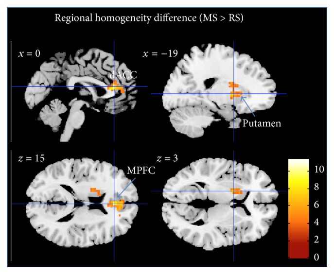
They found that mindfulness training significantly increased the acceptance of splits particularly with the most unfair but profitable splits, increasing from 24% to 44% after training. This resulted in a significant increase in the total distribution of money to both the participant and their supposed partner. Hence, mindfulness training produced greater profit for both. There was no change for the relaxation group. Both groups increased in mindfulness and the greater the increase in mindfulness, the greater the acceptance rates of the offers. In terms of the nervous system activity they found that pre-training unfair offers led to increased activity of the Insula, but this activity was lower after mindfulness training. In addition, in the mindfulness trained participants, cooperation led to increased activity of the Septum. Mindfulness training was also found to increase the functional connectivity between the Insula and the Septum.


These results suggest that mindfulness training produces greater cooperation to the benefit of all participants and that these changes are reflected in changes in neural processing. Why would mindfulness training increase cooperation in the “Ultimatum Game.” Rejecting any off in the game actually reduces the gain for everyone including the participant. So, it is an irrational action. The offers that are rejected are those that are seen to gain more for another than the participant, to be unfair. So, it is this emotional reaction to the perceived unfairness that prompts the counterproductive action of rejecting the offer. Mindfulness training is known to improve emotion regulation, making the individual better able to respond adaptively to emotions. This is probably what made the mindfulness trained group better at cooperation in the face of perceived unfairness, responding rationally and adaptively, increasing profit for all. These effects of mindfulness training are reflected in changes in the brain.


So, build a cooperative brain with mindfulness.


“We are emotional beings and we can’t help but be affected by the varying moods and interactions we have with others. Life is always changing and this constant change can create difficult thoughts and emotions, which can flow into the workplace. The silver lining is that if we can meet suffering at work with concern and care, compassion naturally arises. Work environments that cultivate compassion create a much more positive and productive place to work.” –  Carley Hauck


CMCS – Center for Mindfulness and Contemplative Studies


This and other Contemplative Studies posts are also available on Google+ https://plus.google.com/106784388191201299496/posts


Study Summary

Kirk U, Gu X, Sharp C, Hula A, Fonagy P, Montague PR. Mindfulness training increases cooperative decision making in economic exchanges: Evidence from fMRI. Neuroimage. 2016 Jun 4. pii: S1053-8119(16)30189-6. doi: 10.1016/j.neuroimage.2016.05.075


Abstract

Emotions have been shown to exert influences on decision making during economic exchanges. Here we investigate the underlying neural mechanisms of a training regimen which is hypothesized to promote emotional awareness, specifically mindfulness training (MT). We test the hypothesis that MT increases cooperative economic decision making using fMRI in a randomized longitudinal design involving 8 weeks of either MT or active control training (CT). We find that MT results in an increased willingness to cooperate indexed by higher acceptance rates to unfair monetary offers in the Ultimatum Game. While controlling for acceptance rates of monetary offers between intervention groups, subjects in the MT and CT groups show differential brain activation patterns. Specifically, a subset of more cooperative MT subjects displays increased activation in the septal region, an area linked to social attachment, which may drive the increased willingness to express cooperative behavior in the MT cohort. Furthermore, MT resulted in attenuated activity in anterior insula compared with the CT group in response to unfair monetary offers post-training, which may suggest that MT enables greater ability to effectively regulate the anterior insula and thereby promotes social cooperation. Finally, functional connectivity analyses show a coupling between the septal region and posterior insula in the MT group, suggesting an integration of interoceptive inputs. Together, these results highlight that MT may be employed in contexts where emotional regulation is required to promote social cooperation.


Improve Self-Control and Emotionality with Mindfulness


By John M. de Castro, Ph.D.


“I noticed that people who have addictions and those who teach mindfulness speak the same language. Mindfulness teachers will tell you that stress is caused by craving. If you can let go of that craving, then your stress will dissolve, and practicing mindfulness is the way to do that.” – Judson Brewer


There are many addictions, from exercise, to alcohol, to sex, to drugs, to gambling, to cigarettes. There are a number of differences produced by the specific nature of each addiction. But, there are also some general features. The core components of addiction include an enhanced incentive for the activity or substance (craving), impaired self-control (impulsivity and compulsivity), emotional dysregulation (negative mood) and increased reactivity to stress.


Mindfulness training has been shown to be helpful with each of these components, decreasing cravings, impulsiveness, and psychological and physiological responses to stress, and increasing emotion regulation.  It is no wonder then that mindfulness training has been found to be effective for the treatment of a variety of addictions.  Addictions appear to act via changes to the brain systems affecting self-control; activity in the Anterior Cingulate Cortex (ACC) and adjacent medial Prefrontal Cortex (mPFC). Mindfulness training has also been shown to alter the activity of these structures in the opposite direction through a process called neuroplasticity. This makes a case that mindfulness acts to help in the treatment of addictions by altering the same structures involved in addictions.


In today’s Research News article “Mindfulness meditation improves emotion regulation and reduces drug abuse.” See:

https://www.facebook.com/ContemplativeStudiesCenter/photos/a.628903887133541.1073741828.627681673922429/1279737795383477/?type=3&theater

or below or view the full text of the study at:

http://www.drugandalcoholdependence.com/article/S0376-8716(16)00117-4/fulltext

Tang and colleagues review a number of their studies on the relationships between mindfulness, the psychological properties of addictions, and the neural systems underlying self-control and addiction. They employed a mindfulness meditation training technique called integrative body–mind training (IBMT) and found that it improved executive function, emotions, and responsiveness to stress.


In other studies, they investigated the brain’s response to IBMT and found that it increased activity in the Anterior Cingulate Cortex (ACC) which is known to be involved in emotion regulation and the activity of the parasympathetic nervous system, that is known to be an antidote to stress responses. These results strongly suggest that mindfulness training (IBMT) acts in ways that would tend to counteract the effects of addictions including self-control and stress responsiveness. Tang and colleagues went on to test IBMT on students who were addicted to cigarettes (nicotine). They found that smokers had lower activity in Anterior Cingulate Cortex (ACC) and the medial Prefrontal Cortex (mPFC). But, IBMT training significantly reduced cigarette smoking and increased both ACC and mPFC activity.


These results suggest that mindfulness meditation increases activity in ACC and mPFC which are involved in emotion regulation and self-control. The improvements in these areas, in turn, assist in the prevention and treatment of addictions. These studies have produced an integrated theory of how mindfulness effects addiction by altering the nervous system in such a way as to counteract the psychological issues that underlie addictions. These studies are leading to a better understanding of how mindfulness training produces improvements in addictions.


So, improve self-control and emotionality with mindfulness and reduce addictions.


“mindfulness is likely an effective tool in helping people with addiction because it’s a single, simple skill that a person can practice multiple times throughout their day, every day, regardless of the life challenges that arise. With so much opportunity for practice—rather than, say, only practicing when someone offers them a cigarette—people can learn that skill deeply.” –  James Davis


CMCS – Center for Mindfulness and Contemplative Studies


This and other Contemplative Studies posts are also available on Google+ https://plus.google.com/106784388191201299496/posts


Study Summary

Tang YY, Tang R, Posner MI. Mindfulness meditation improves emotion regulation and reduces drug abuse. Drug Alcohol Depend. 2016 Jun 1;163 Suppl 1:S13-8. doi: 10.1016/j.drugalcdep.2015.11.041


Highlights

  • We review the effects of mindfulness meditation on emotion regulation and addiction.
  • We propose the brain mechanism of mindfulness meditation.
  • We examine addiction treatment using mindfulness meditation.

Abstract

BACKGROUND: The core clinical symptoms of addiction include an enhanced incentive for drug taking (craving), impaired self-control (impulsivity and compulsivity), emotional dysregulation (negative mood) and increased stress reactivity. Symptoms related to impaired self-control involve reduced activity in anterior cingulate cortex (ACC), adjacent prefrontal cortex (mPFC) and other brain areas. Behavioral training such asmindfulness meditation can increase the function of control networks including those leading to improved emotion regulation and thus may be a promising approach for the treatment of addiction.

METHODS: In a series of randomized controlled trials (RCTs), we tested whether increased ACC/mPFC activity is related to better self-control abilities in executive functions, emotion regulation and stress response in healthy and addicted populations. After a brief mindfulness training (Integrative Body-Mind Training, IBMT), we used the Positive and Negative Affect Schedule (PANAS) and Profile of Mood States (POMS) to measure emotion regulation, salivary cortisol for the stress response and fMRI for brain functional and DTI structural changes. Relaxation training was used to serve as an active control.

RESULTS: In both smokers and nonsmokers, improved self-control abilities in emotion regulation and stress reduction were found after training and these changes were related to increased ACC/mPFC activity following training. Compared with nonsmokers, smokers showed reduced ACC/mPFC activity in the self-control network before training, and these deficits were ameliorated after training.

CONCLUSIONS: These results indicate that promoting emotion regulation and improving ACC/mPFC brain activity can help for addiction prevention and treatment.

http://www.drugandalcoholdependence.com/article/S0376-8716(16)00117-4/fulltext


Awakening-like Experiences Occur with Epilepsy in the Insula

mystical experiences epilepsy2 Gschwing

By John M. de Castro, Ph.D.


“Dostoevsky described his seizures in a letter to a friend: “I feel entirely in harmony with myself and the whole world, and this feeling is so strong and so delightful that for a few seconds of such bliss one would gladly give up 10 years of one’s life, if not one’s whole life.”” – Anil Ananthaswamy


Millions of people worldwide perform practices, such as meditation, yoga, and prayer, to achieve a spiritual awakening. Others use drugs such as peyote, ayahuasca  and psilocybin to induce spiritual awakenings. If successful, these people report unique profound experiences that permanently alter their lives and the way they perceive the world. These experiences have many characteristics many of which are unique to the experiencer, their religious context, and their present situation. They include a state of ecstasy, bliss, love and joy, a sense of ultimate freedom and belonging, a transcendence of space and time, a sense of lacking control over the event, a greater sense of meaning and purpose of life, a sense of timelessness, a sense of having encountered ultimate reality, a sense of sacredness, a sense that one cannot adequately describe the richness of this experience. But, the common, central feature of all of these experiences is a sense on oneness, that all things are contained in a single thing, a sense of union with the universe and/or God and everything in existence.


The fact that these experiences can be induced by drugs and that drugs have their effects by altering the chemistry of the nervous system, has led to the notion that perhaps these experiences are not actually spiritual but rather simply an altered state of the brain produced by drugs or intense spiritual practices. An important observation in this regard is that alterations of the brain can make it more likely that an individual will have a spiritual experience. Spiritual experiences can occur occasionally with epileptic seizures. This may provide clues as to what neural structures are involved in spiritual experiences.


In today’s Research News article “Ecstatic Epileptic Seizures: A Glimpse into the Multiple Roles of the Insula.” See:

https://www.facebook.com/ContemplativeStudiesCenter/photos/a.628903887133541.1073741828.627681673922429/1278188525538404/?type=3&theater

or below or view the full text of the study at:

http://www.ncbi.nlm.nih.gov/pmc/articles/PMC4756129/

Gschwind and Picard review published cases of individuals who report spiritual experiences with the onset of an epileptic seizure. Their review led them to hypothesize that the focal area for the production of spiritual experiences is the insular cortex. This is a large piece pf cerebral cortex that has been enfolded with growth of the cortex and does not appear on the surface. It has been covered and is buried deep inside at the juncture of the parietal, temporal, and frontal lobes. The insula is highly interconnected with a wide variety of other cortical and subcortical areas of the brain. It has been implicated in consciousness and appears to play a role in diverse functions usually linked to emotion or the regulation of the body’s homeostasis. These functions include perception, motor control, self-awareness, cognitive functioning, and interpersonal experience. So, the insula would appear to be well situated to produce and affect higher level experiences.


Gschwind and Picard review cases of spiritual (ecstatic) experiences that occur with epileptic seizures either located in or affecting the activity of the insula. In addition, they review cases where direct electrical stimulation of the insula produce reports of spiritual-like experiences in awake humans. This together with the widespread interconnectivity of the insula and its suspected role in higher conscious experience, makes a case that the insula is the area of the brain that is central to spiritual (ecstatic) experiences.


The reviewed information is highly complex and the conclusions are speculative. They are far from proving the case that the insula is responsible for spiritual (ecstatic) experiences. But, it provides sufficient enticing evidence that further scrutiny of this area and its association to these experiences should be undertaken. It should be mentioned that no area in the brain works alone. Rather complex behaviors and experiences are produced by the joint action of large numbers of areas throughout the brain. So, pointing to the insula only suggests that it may be central to the disparate neural system underlying spiritual (ecstatic) experiences.


That spiritual (ecstatic) experiences can be produced by epileptic seizures and by electrical stimulation of the brain, combined with the fact that drugs that alter brain chemistry can also produce these experiences, suggests that neural systems may underlie all spiritual experiences. This should not be surprising as the experiences are physical in nature, very unusual sensory experiences, but physical experiences nonetheless. So, it should be as no surprise that a physical entity, the brain, may underlie them. It has yet to be conclusively shown, however, the experiences produced by epilepsy, stimulation, or drugs is the same as that reported by mystics and spiritual seekers. This also leaves open the question as to what it is that’s registering and aware of. these experiences. It appears that regardless of what produces them, the underlying awareness registering them is unchanged.


“The anterior insula may be where all manner of feelings, including bodily pleasures and pains; sights, sounds and smells; emotions, expectations and intentions; and even the sense of one’s physical surroundings are integrated into a unified sense of a “self” moving through time.

If this hypothesis is true, then disturbances to the anterior insula would likely cause disturbances to the usual sense of self-awareness.Jim Schnabel


CMCS – Center for Mindfulness and Contemplative Studies


This and other Contemplative Studies posts are available at the Contemplative Studies Blog http://contemplative-studies.org/wp/


Study Summary

Gschwind, M., & Picard, F. (2016). Ecstatic Epileptic Seizures: A Glimpse into the Multiple Roles of the Insula. Frontiers in Behavioral Neuroscience, 10, 21. http://doi.org/10.3389/fnbeh.2016.00021


Abstract

Ecstatic epileptic seizures are a rare but compelling epileptic entity. During the first seconds of these seizures, ecstatic auras provoke feelings of well-being, intense serenity, bliss, and “enhanced self-awareness.” They are associated with the impression of time dilation, and can be described as a mystic experience by some patients. The functional neuroanatomy of ecstatic seizures is still debated. During recent years several patients presenting with ecstatic auras have been reported by others and us (in total n = 52); a few of them in the setting of presurgical evaluation including electrical brain stimulation. According to the recently recognized functions of the insula, and the results of nuclear brain imaging and electrical stimulation, the ecstatic symptoms in these patients seem to localize to a functional network centered around the anterior insular cortex, where we thus propose to locate this rare ictal phenomenon. Here we summarize the role of the multiple sensory, autonomic, affective, and cognitive functions of the insular cortex, which are integrated into the creation of self-awareness, and we suggest how this system may become dysfunctional on several levels during ecstatic aura.


Be Less Controlled by Rewards with Mindfulness

By John M. de Castro, Ph.D.


“Mindfulness should no longer be considered a “nice-to-have.” It’s a “must-have”:  a way to keep our brains healthy, to support self-regulation and effective decision-making capabilities, and to protect ourselves from toxic stress. It can be integrated into one’s religious or spiritual life, or practiced as a form of secular mental training.  When we take a seat, take a breath, and commit to being mindful, particularly when we gather with others who are doing the same, we have the potential to be changed.” – Christina Congleton


Behavioral Psychology teaches that we are slaves to rewards and punishers. The Law of Effect states that we tend to repeat actions that are followed by a pleasing state of affairs (rewards) and are less likely to repeat actions that are followed by an aversive state of affairs (punishers). It is evident that people respond this way. It can be healthy and adaptive as long as it is kept under reasonable restraints and at moderate levels.


People who are too attached to rewards such as money are never really satisfied no matter how much they acquire. They are on what is called the “hedonic treadmill” where reward produces brief happiness but once it diminishes the individual becomes unhappy and works even harder for more reward and on and on it goes.  On the other hand, a lack of responsiveness to rewards and punishers is a hallmark of depression. But, the ability to delay reward is a characteristic of very successful people. It has been shown that, demonstrating the ability in early childhood to postpone reward to get a bigger reward later, predicts success in adult life.


So, it is crucial for well-being that we learn moderation and control of our responses to rewards and punishers. Mindfulness training has been shown to assist in restraining impulsivity and improving response inhibition and delay of gratification. In today’s Research News article “Adaptive neural reward processing during anticipation and receipt of monetary rewards in mindfulness meditators.” See:

https://www.facebook.com/ContemplativeStudiesCenter/photos/a.628903887133541.1073741828.627681673922429/1257081990982391/?type=3&theater

or below or view the full text of the study at:

http://www.ncbi.nlm.nih.gov/pmc/articles/PMC4420752/

Kirk and colleagues investigate the effect of mindfulness on reward delay and the neural circuits involved. They compared the neural responses of experienced meditators vs. meditation naive participants to a monetary incentive delay task. To be successful in this task in earning money or preventing monetary loss the participant has to withhold responding until signaled. During the performance of this task the participants had their brain activity recorded with magnetic resonance imaging (f-MRI).


They did not find a difference in the groups in performance of the monetary incentive delay task. There were, however, significant differences in brain activation between the groups. In anticipation of rewards the experienced meditators had significantly reduced activity in the dorsal striatum and increased activity in the posterior insula. When the experienced meditators received a reward they had significantly less activation of the Ventromedial Prefrontal Cortex than the controls.


The results suggest that the experienced meditators are less activated during reward anticipation in the striatum which has been shown to be activated during reward anticipation. This suggests that the meditators have a moderated response to the thought of attaining a reward. They also appear to be more in touch with their internal feelings during reward anticipation as they had increased activation during reward anticipation of the posterior insula which is known to be involved in the processing of internal sensations. The experienced meditators also appear to have a reduced valuation of the reward during reward receipt as suggested by the reduced activation of the Ventromedial Prefrontal Cortex that has been shown to be involved in valuing reward magnitude.


The results thus suggest that meditation practice alters the brain producing more moderate response to the anticipation and presentation of reward and producing greater sensitivity to their internal state. Both of these modifications would be expected to make meditators better at keeping rewards and punishers in perspective, not being taken away on the “hedonic treadmill,” and being better able to delay gratification. This would in turn predict greater success in life as a results of meditation practice.


So, be less controlled by rewards with mindfulness.


“Meditation gives you the wherewithal to pause, observe how easily the mind can exaggerate the severity of a setback, and resist getting drawn back into the abyss.” — Richie Davidson


CMCS – Center for Mindfulness and Contemplative Studies


This and other Contemplative Studies posts are also available on Google+ https://plus.google.com/106784388191201299496/posts


Study Summary

Kirk, U., Brown, K. W., & Downar, J. (2015). Adaptive neural reward processing during anticipation and receipt of monetary rewards in mindfulness meditators. Social Cognitive and Affective Neuroscience, 10(5), 752–759. http://doi.org/10.1093/scan/nsu112


Abstract

Reward seeking is ubiquitous and adaptive in humans. But excessive reward seeking behavior, such as chasing monetary rewards, may lead to diminished subjective well-being. This study examined whether individuals trained in mindfulness meditation show neural evidence of lower susceptibility to monetary rewards. Seventy-eight participants (34 meditators, 44 matched controls) completed the monetary incentive delay task while undergoing functional magnetic resonance imaging. The groups performed equally on the task, but meditators showed lower neural activations in the caudate nucleus during reward anticipation, and elevated bilateral posterior insula activation during reward anticipation. Meditators also evidenced reduced activations in the ventromedial prefrontal cortex during reward receipt compared with controls. Connectivity parameters between the right caudate and bilateral anterior insula were attenuated in meditators during incentive anticipation. In summary, brain regions involved in reward processing—both during reward anticipation and receipt of reward—responded differently in mindfulness meditators than in nonmeditators, indicating that the former are less susceptible to monetary incentives.

http://www.ncbi.nlm.nih.gov/pmc/articles/PMC4420752/


Tone Down the Ringing in the Ears with Mindfulness

By John M. de Castro, Ph.D.


“Mindful meditation helped me to think (and not think) about tinnitus in ways that had not occurred before, and in doing so made tinnitus much less of a burden to carry. It did not fix my tinnitus but it fixed me in a way that made tinnitus easier to bear. I now feel as if I live in the same universe as everybody else. I wouldn’t be anywhere else.” – Claire Bartlett


Tinnitus is one of the most common symptoms to affect humanity. People with tinnitus live with a phantom noise that can range from a low hiss or ringing to a loud roar or squeal which can be present constantly or intermittently. It can have a significant impact on people’s ability to hear, concentrate, or even participate in everyday activities. Tinnitus is not a disease itself; it is a symptom that something is wrong in the auditory system. The vast majority of people with tinnitus have what is known as subjective tinnitus. This is caused by unknown problems somewhere in the auditory system; the inner, middle, or outer ear, the part of the brain that translates nerve signals as sounds, or the auditory nerves.


Approximately 25 million to 50 million people in the United States experience it to some degree. Tinnitus and hearing loss are the top service-related disabilities among veterans; 60 percent of those who served in Iraq and Afghanistan return home with hearing loss. Approximately 16 million people seek medical attention for their tinnitus, and for up to two million patients, debilitating tinnitus interferes with their daily lives. Tinnitus is sometimes the first sign of hearing loss, occurring in roughly 90 percent of tinnitus cases. There are a number of treatments for tinnitus including, counseling, sound therapy, drugs, and even brain stimulation. Unfortunately, none of these treatments is very effective. Hence there is a need for safe and effective alternative treatments for tinnitus.


In today’s Research News article “Effects of Mindfulness-Based Stress Reduction Therapy On Subjective Bother and Neural Connectivity in Chronic Tinnitus.” See:

https://www.facebook.com/ContemplativeStudiesCenter/photos/a.628903887133541.1073741828.627681673922429/1250875274936396/?type=3&theater

or below or view the full text of the study at:

http://www.ncbi.nlm.nih.gov/pmc/articles/PMC4650869/

Roland and colleagues investigate Mindfulness Based Stress Reduction (MBSR) training as a treatment for tinnitus. MBSR is an 8-week program that includes meditation, yoga, and body scan techniques. There are once a week 2-hour meetings and daily home practice. They recruited adult tinnitus sufferers, measured symptoms and scanned their brains with Magnetic Resonance Imaging (MRI) for brain structure and functional connectivity. Subsequently they received MBSR training followed by tinnitus measurements and MRI scanning.


They found a clinically significant decrease in both tinnitus symptoms and the degree of handicap produced by tinnitus after MBSR that was maintained at a one month follow up examination. They also found that after MBSR training there was increased functional connectivity with the prefrontal and operculum cortexes which are known to be associated with attention mechanisms, but no change in the default mode network that is associated with mind wandering and self-referential thoughts. These connectivity results make sense as MBSR is targeted at improving attention to the body and the present moment.


These results are very exciting as they suggest that Mindfulness Based Stress Reduction (MBSR) training may be a safe and effective treatment for tinnitus. The symptom improvements were substantial and over 60% of the tinnitus sufferers had clinically significant improvements. They further suggest that MBSR may improve tinnitus symptoms by increasing the effectiveness of brain attentional networks. It is possible that, by improving attention to the present moment, MBSR results in less attention being paid to the tinnitus, reducing its impact.


It should be kept in mind, however, that this was a pilot study without a control group. Hence the conclusions must be tempered with caution until a definitive randomized controlled trial can be conducted. The results are encouraging enough that such a trial is warranted.


Nevertheless, tone down the ringing in the ears with mindfulness.


“participants commented that tinnitus no longer seemed like a dreadful curse; it was just another sensation that could be annoying but was not insurmountable.” – Jennifer Gans


CMCS – Center for Mindfulness and Contemplative Studies


This and other Contemplative Studies posts are available on Google+ https://plus.google.com/106784388191201299496/posts


Study Summary

Roland, L. T., Lenze, E. J., Hardin, F. M., Kallogjeri, D., Nicklaus, J., Wineland, A., … Piccirillo, J. F. (2015). Effects of Mindfulness-Based Stress Reduction Therapy On Subjective Bother And Neural Connectivity In Chronic Tinnitus. Otolaryngology–Head and Neck Surgery : Official Journal of American Academy of Otolaryngology-Head and Neck Surgery, 152(5), 919–926. http://doi.org/10.1177/0194599815571556


Abstract

Objective: To evaluate the impact of an MBSR program in patients with chronic bothersome tinnitus on the 1) severity of symptoms of tinnitus and 2) functional connectivity in neural attention networks.

Study Design

Open-label interventional pilot study.

Setting: Outpatient academic medical center.

Subjects: A total of 13 adult participants with a median age of 55 years, suffering from bothersome tinnitus.

Methods: An 8-week MBSR program was conducted by a trained MBSR instructor. The primary outcome measure was the difference in patient-reported tinnitus symptoms using the Tinnitus Handicap Index (THI) andTinnitus Functional Index (TFI) between pre-intervention, post-MBSR, and 4-week post-MBSR assessments. Secondary outcomes included change in measurements of depression, anxiety, mindfulness and cognitive abilities. Functional connectivity MRI was performed at pre- and post- MBSR intervention time points to serve as a neuroimaging biomarker of critical cortical networks.

Results: Scores on the THI and TFI showed statistically significant and clinically meaningful improvement over the course of the study with a median ΔTHI of −16 and median ΔTFI of −14.8 between baseline and 4-week follow-up scores. Except for depression, there was no significant change in any of the secondary outcome measures. Analysis of the rs-fcMRI data showed increased connectivity in the post-MBSR group in attention networks but not the default network.

Conclusion: Participation in an MBSR program is associated with decreased severity in tinnitus symptoms and depression, and connectivity changes in neural attention networks. MBSR is a promising treatment option for chronic bothersome tinnitus that is both noninvasive and inexpensive.

http://www.ncbi.nlm.nih.gov/pmc/articles/PMC4650869/


Increase Hemispheric Blood Oxygenation with Yogic Nostril Breathing


By John M. de Castro, Ph.D.


“Breathing in through your left nostril will access the right “feeling” hemisphere of your brain, and breathing in through your right nostril, will access the left “thinking” hemisphere of your brain.  Consciously alternating your breath between either nostril will allow you to activate and access your whole brain.” – Carole Bourne


Mindfulness practices have been shown to alter the brain, including short-term changes in activity and longer-term changes in the size and connectivity of brain areas and the chemistry of the nervous system. It is thought that many of the beneficial effects of mindfulness practices are mediated by these changes in the nervous system. Yoga practice is a mindfulness technique that has documented benefits for the individual’s psychological and physical health and well-being. It has also been shown to produce short- and long-term changes in the brain. It is thus likely that yoga practice also produces its beneficial effects by altering the brain.


Yoga consists of a number of components including, poses, breathing exercises, meditation, concentration, and philosophy/ethics.  So, it is difficult to determine which facet or combination of facets of yoga are responsible for which benefit. Hence, it is important to begin to test each component in isolation to determine its effects. In a previously reviewed study the effects of yogic breathing techniques, specifically, left or right or alternating nostril breathing, it was found that these forms of breathing produce improvements in spatial and verbal memory ability. (LINK to Garg et al. study) These effects of left or right nostril breathing are thought to be produced by changing the oxygen flows to the individual neural hemispheres.


In today’s Research News article “Effect of uninostril yoga breathing on brain hemodynamics: A functional near-infrared spectroscopy study.” See:

https://www.facebook.com/ContemplativeStudiesCenter/photos/a.628903887133541.1073741828.627681673922429/1250194438337813/?type=3&theater

or below or view the full text of the study at: http://www.ncbi.nlm.nih.gov/pmc/articles/PMC4728953/

Singh and colleagues examine the effects of left or right nostril breathing on oxygen and blood volume flows to the left and right hemispheres. They measured blood volume and oxygen levels in experienced yoga practitioners using a sophisticated optical imaging technique called Functional near-infrared spectroscopy. They were measured on separate days for the effects of left nostril, right nostril, or breath awareness (control condition) on hemispheric blood flows.


They found that with right nostril breathing there was an increase in blood oxygen and blood volume to the left prefrontal cortex while left nostril breathing produced an increase in blood oxygen and blood volume to the right prefrontal cortex. Hence, nostril breathing produces increased flow and oxygenation to the contralateral hemisphere. This would suggest that right nostril breathing would produce increments in left hemisphere functions such as verbal, mathematical, or logical functions, while left nostril breathing would produce increases in right hemisphere functions such as spatial, emotional, or artistic functions. It will be up to future research to test this conjecture.


The results, though, do demonstrate that yogic nostril breathing differentially effects blood flows and oxygenation to the contralateral hemisphere. This provides an underlying mechanism for the effects of yogic nostril breathing.


So, increase hemispheric blood oxygenation with yogic nostril breathing.


CMCS – Center for Mindfulness and Contemplative Studies


“Breathing consciously is a powerful act. The process of breathing sits directly at the interface of our voluntary nervous system (aspects of our physiology under our conscious control) and our autonomic nervous system (aspects generally not under conscious control). It’s a direct path for us to communicate quickly to the brain via what we do with our body. – Paula Watkins


This and other Contemplative Studies posts are available on Google+ https://plus.google.com/106784388191201299496/posts


Study Summary

Singh, K., Bhargav, H., & Srinivasan, T. (2016). Effect of uninostril yoga breathing on brain hemodynamics: A functional near-infrared spectroscopy study. International Journal of Yoga, 9(1), 12–19. http://doi.org/10.4103/0973-6131.171711


Abstract

Objectives: To measure the effect of the right and left nostril yoga breathing on frontal hemodynamic responses in 32 right handed healthy male subjects within the age range of 18–35 years (23.75 ± 4.14 years).

Materials and Methods: Each subject practiced right nostril yoga breathing (RNYB), left nostril yoga breathing (LNYB) or breath awareness (BA) (as control) for 10 min at the same time of the day for three consecutive days, respectively. The sequence of intervention was assigned randomly. The frontal hemodynamic response in terms of changes in the oxygenated hemoglobin (oxyHb), deoxygenated hemoglobin (deoxyHb), and total hemoglobin (totalHb or blood volume) concentration was tapped for 5 min before (pre) and 10 min during the breathing practices using a 16 channel functional near-infrared system (FNIR100-ACK-W, BIOPAC Systems, Inc., U.S.A.). Average of the eight channels on each side (right and left frontals) was obtained for the two sessions (pre and during). Data was analyzed using SPSS version 10.0 through paired and independent samples t-test.

Results: Within group comparison showed that during RNYB, oxyHb levels increased significantly in the left prefrontal cortex (PFC) as compared to the baseline (P = 0.026). LNYB showed a trend towards significance for reduction in oxyHb in the right hemisphere (P = 0.057). Whereas BA caused significant reduction in deoxyHb (P = 0.023) in the left hemisphere. Between groups comparison revealed that oxyHb and blood volume in the left PFC increased significantly during RNYB as compared to BA (oxyHb: P =0.012; TotalHb: P =0.017) and LNYB (oxyHb: P =0.024; totalHb: P =0.034).

Conclusion: RNYB increased oxygenation and blood volume in the left PFC as compared to BA and LNYB. This supports the relationship between nasal cycle and ultradian rhythm of cerebral dominance and suggests a possible application of uninostril yoga breathing in the management of psychopathological states which show lateralized cerebral dysfunctions.

http://www.ncbi.nlm.nih.gov/pmc/articles/PMC4728953/


Change the Traumatized Brain with Mindfulness


By John M. de Castro, Ph.D.


“Mind-body exercise offers a low-cost approach that could be used as a complement to traditional psychotherapy or drug treatments. These self-directed practices give PTSD patients control over their own treatment and have few side effects.” – Sang H. Kim


Experiencing trauma is quite common. It has been estimated that 60% of men and 50% of women will experience a significant traumatic event during their lifetime. But, only a fraction will develop Post-Traumatic Stress Disorder (PTSD). But this still results in a frightening number of people with 7%-8% of the population developing PTSD at some point in their life. For military personnel it’s much more likely for PTSD to develop with about 11%-20% of those who have served in a war zone developing PTSD.


PTSD involves a number of troubling symptoms including reliving the event with the same fear and horror in nightmares or with a flashback. PTSD sufferers avoid situations that remind them of the event this may include crowds, driving, movies, etc. and may avoid seeking help because it keeps them from having to think or talk about the event. They often experience negative changes in beliefs and feelings including difficulty experiencing positive or loving feelings toward other people, avoiding relationships, memory difficulties, or see the world as dangerous and no one can be trusted. Sufferers may feel hyperarousal, feeling keyed up and jittery, or always alert and on the lookout for danger. They may experience sudden anger or irritability, may have a hard time sleeping or concentrating, may be startled by a loud noise or surprise.


Obviously, these are troubling symptoms that need to be addressed. There are a number of therapies that have been developed to treat PTSD. One of which, mindfulness training has been found to be particularly effective. The nervous system is constantly changing and adapting to the environment. It will change size, activity, and connectivity in response to experience. For example, the brain area that controls the right index finger has been found to be larger in blind subjects who use braille than in sighted individuals.  Similarly, cab drivers in London who navigate the twisting streets of the city, have a larger hippocampus, which is involved in spatial navigation, than predefined route bus drivers. These changes in the brain are called neuroplasticity. Over the last decade neuroscience has been studying the effects of contemplative practices on the brain and has identified neuroplastic changes in widespread areas. This all suggests that mindfulness training would improve PTSD symptoms by producing changes to the brains of sufferers.


In today’s Research News article “Altered default mode network (DMN) resting state functional connectivity following a mindfulness-based exposure therapy for posttraumatic stress disorder (PTSD) in combat veterans of Afghanistan and Iraq.” See:

https://www.facebook.com/ContemplativeStudiesCenter/photos/a.628903887133541.1073741828.627681673922429/1223008144389776/?type=3&theater

or below

King and colleagues perform functional Magnetic Resonance Imaging (f-MRI) before and after a 16-week mindfulness treatment program for Post-Traumatic Stress Disorder (PTSD) in veterans and compared the results to veterans treated with a present-centered group therapy for PTSD. They found that both groups showed improvement in PTSD symptoms, but only the mindfulness treatment produced significant improvement in total and intrusive, avoidant, and hyperarousal PTSD symptoms. This replicates previous results that mindfulness training is effective in treating PTSD symptoms.


King and colleagues also found that the mindfulness treatment produced changes in the nervous system. In particular, they found increased connectivity between the dorsolateral prefrontal cortex and dorsal anterior cingulate cortex. These are structures included in what has been termed the Default Mode Network (DMN) which is involved in producing mind wandering and intrusive thoughts. In addition, they found that the greater the increase in connectivity of these structures the greater the improvement in PTSD symptoms. These results suggest that mindfulness training changes the nervous system in such a way as to improve the ability of the individual to shift attention away from mind wandering and intrusive thoughts. These off-task thoughts are characteristic of PTSD sufferers and are a key symptom of the disorder. By being able to shift away from these thoughts and pay attention to the present moment, the mindfulness trained individuals are better able to keep these thoughts from tormenting them and thereby improve PTSD.


These results demonstrate the effectiveness of mindfulness training for Post-Traumatic Stress Disorder (PTSD) and suggest a mechanism by which the training produces these effects. It appears to alter the Default Mode Network (DMN) of the brain allowing the individual to better pay attention to the present moment and shift thinking away from the intrusive thoughts like flashbacks that are characteristic of PTSD.


So, change the traumatized brain with mindfulness.


“Very often, depression can be about the past, ruminating over and over about losses or other terrible things that happened in the past. Anxiety, meanwhile, very often involves ruminating about terrible things that you’re afraid are going to happen in the future. But in the present, very often, there’s actually nothing terrible going on, and the act of recognizing that can be helpful.” – Anthony King


CMCS – Center for Mindfulness and Contemplative Studies


Study Summary

King, A. P., Block, S. R., Sripada, R. K., Rauch, S., Giardino, N., Favorite, T., Angstadt, M., Kessler, D., Welsh, R. and Liberzon, I. (2016), Altered default mode network (DMN) resting state functional connectivity following a mindfulness-based exposure therapy for posttraumatic stress disorder (PTSD) in combat veterans of Afghanistan and Iraq. Depress. Anxiety, 33: 289–299. doi: 10.1002/da.22481


Abstract

Background: Recent studies suggest that mindfulness may be an effective component for posttraumatic stress disorder (PTSD) treatment. Mindfulness involves practice in volitional shifting of attention from “mind wandering” to present-moment attention to sensations, and cultivating acceptance. We examined potential neural correlates of mindfulness training using a novel group therapy (mindfulness-based exposure therapy (MBET)) in combat veterans with PTSD deployed to Afghanistan (OEF) and/or Iraq (OIF).

Methods: Twenty-three male OEF/OIF combat veterans with PTSD were treated with a mindfulness-based intervention (N = 14) or an active control group therapy (present-centered group therapy (PCGT), N = 9). Pre-post therapy functional magnetic resonance imaging (fMRI, 3 T) examined resting-state functional connectivity (rsFC) in default mode network (DMN) using posterior cingulate cortex (PCC) and ventral medial prefrontal cortex (vmPFC) seeds, and salience network (SN) with anatomical amygdala seeds. PTSD symptoms were assessed at pre- and posttherapy with Clinician Administered PTSD Scale (CAPS).

Results: Patients treated with MBET had reduced PTSD symptoms (effect size d = 0.92) but effect was not significantly different from PCGT (d = 0.46). Increased DMN rsFC (PCC seed) with dorsolateral dorsolateral prefrontal cortex (DLPFC) regions and dorsal anterior cingulate cortex (ACC) regions associated with executive control was seen following MBET. A group × time interaction found MBET showed increased connectivity with DLPFC and dorsal ACC following therapy; PCC–DLPFC connectivity was correlated with improvement in PTSD avoidant and hyperarousal symptoms.

Conclusions: Increased connectivity between DMN and executive control regions following mindfulness training could underlie increased capacity for volitional shifting of attention. The increased PCC–DLPFC rsFC following MBET was related to PTSD symptom improvement, pointing to a potential therapeutic mechanism of mindfulness-based therapies.


Change the Depressed Brain with Meditation

Meditation brain depression2 Yang

By John M. de Castro, Ph.D.


“Meditation isn’t a magic bullet for depression, as no treatment is, but it’s one of the tools that may help manage symptoms.” – Alice Walton


Depression is the most common mental illness affecting over 6% of the population.  It is debilitating by producing any or all of a long list of symptoms including: feelings of sadness or unhappiness, change in appetite or weight, slowed thinking or speech, loss of interest in activities or social gatherings, fatigue, loss in energy, sleeplessness, feelings of guilt or anger over past failures, trouble concentrating, indecisiveness, anger or frustration for no distinct reason, thoughts of dying, death and suicide. The first line treatment is antidepressant drugs. But, depression can be difficult to treat. Of patients treated initially with drugs only about a third attained remission of the depression. After repeated and varied treatments including drugs, therapy, exercise etc. only about two thirds of patients attained remission. This leaves a third of all patients treated without success.


Mindfulness practices have been demonstrated to be beneficial for depression. They not only reduce depression levels in relatively normal people but also relieve depression in individual with major depression. They can even help in cases where drugs fail to relieve the depression. In addition, mindfulness practices can reduce the likelihood of a relapse after successful treatment for depression. In other words, mindfulness is an effective treatment either alone or in combination with drugs for depression.


In the last few decades it has become increasingly clear that the brain is very plastic and can be reshaped by what we do and what we experience. This has been called neuroplasticity. Contemplative practices have been shown to produce neuroplastic change altering the brain. They tend to increase the size, activity, and connectivity of areas of the brain that are important for attention, and emotion regulation while reducing the size, activity, and connectivity of areas of the brain that are involved in mind wandering and self-centered thinking, daydreaming, and rumination.


Mindfulness practices appear to take advantage of neuroplasticity to act upon the chemistry of the brain and brain electrical activity to help relieve depression. Hence, it makes sense that there should be further research on the effects of mindfulness on the brain and depression to better understand the mechanisms of action of mindfulness and potentially optimize treatment. In today’s Research News article “State and Training Effects of Mindfulness Meditation on Brain Networks Reflect Neuronal Mechanisms of Its Antidepressant Effect”

https://www.facebook.com/ContemplativeStudiesCenter/photos/a.628903887133541.1073741828.627681673922429/1217282338295690/?type=3&theater

http://www.ncbi.nlm.nih.gov/pmc/articles/PMC4779536/

Yang and colleagues trained meditation naive college students for 8-weeks with a mindfulness program based upon a combination of Mindfulness Based Stress Reduction (MBSR) and Acceptance and Commitment Therapy (ACT). Functional Magnetic Resonance Imaging (f-MRI) brain scans were performed before training and 40 days later both at rest and during meditation. The students emotional state and depression levels were also measured before and after training.


After mindfulness training there was a 45% reduction in depression, a 21% reduction in trait anxiety, and a small reduction in tension. The brain was also found to be changed at rest after training. The changes were many and diverse. But arguably the most interesting change was a reduction in the functional connectivity between the pregenual anterior cingulate and dorsal medical prefrontal cortex. The anterior cingulate cortex is part of what is called the default mode network that is activated during mind wandering and emotionality. It is involved in major depression and the loss of emotionality, so called flat affect, that characterizes depression. On the other hand, the dorsal medical prefrontal cortex is involved in focused attention, cognitive control, and emotion regulation. These results are very complex and must be interpreted cautiously. But, it appears that meditation training reduces the ability of areas involved in depression from affecting areas involved in thought and emotion. This is a potential route for mindfulness affects in relieving depression.


Regardless, it’s clear that mindfulness can change the depressed brain.


“We can intentionally shape the direction of plasticity changes in our brain. By focusing on wholesome thoughts, for example, and directing our intentions in those ways, we can potentially influence the plasticity of our brains and shape them in ways that can be beneficial.” – Richie Davidson


CMCS – Center for Mindfulness and Contemplative Studies


Study Summary


Yang, C.-C., Barrós-Loscertales, A., Pinazo, D., Ventura-Campos, N., Borchardt, V., Bustamante, J.-C., … Walter, M. (2016). State and Training Effects of Mindfulness Meditation on Brain Networks Reflect Neuronal Mechanisms of Its Antidepressant Effect. Neural Plasticity, 2016, 9504642. http://doi.org/10.1155/2016/9504642


Abstract

The topic of investigating how mindfulness meditation training can have antidepressant effects via plastic changes in both resting state and meditation state brain activity is important in the rapidly emerging field of neuroplasticity. In the present study, we used a longitudinal design investigating resting state fMRI both before and after 40 days of meditation training in 13 novices. After training, we compared differences in network connectivity between rest and meditation using common resting state functional connectivity methods. Interregional methods were paired with local measures such as Regional Homogeneity. As expected, significant differences in functional connectivity both between states (rest versus meditation) and between time points (before versus after training) were observed. During meditation, the internal consistency in the precuneus and the temporoparietal junction increased, while the internal consistency of frontal brain regions decreased. A follow-up analysis of regional connectivity of the dorsal anterior cingulate cortex further revealed reduced connectivity with anterior insula during meditation. After meditation training, reduced resting state functional connectivity between the pregenual anterior cingulate and dorsal medical prefrontal cortex was observed. Most importantly, significantly reduced depression/anxiety scores were observed after training. Hence, these findings suggest that mindfulness meditation might be of therapeutic use by inducing plasticity related network changes altering the neuronal basis of affective disorders such as depression.

http://www.ncbi.nlm.nih.gov/pmc/articles/PMC4779536/


Healthy Aging – Improve the Brain and Memory with Tai Chi

Healthy Aging – Improve the Brain and Memory with Tai Chi


By John M. de Castro, Ph.D.


“Another great benefit of Tai Chi is that it’s accessible to people of all ages and fitness abilities. It’s the focus on the subtle movements that exercise the brain and boost cognitive abilities. First, it’s learning the precise movements of the ancient martial art form that will give your brain a boost. Then, it’s the continued focus linking the breath and the movements.” – Karl Romain


We all want to live longer. We celebrate the increasing longevity of the population. But, aging is a mixed blessing. The aging process involves a systematic progressive decline in every system in the body, the brain included. It cannot be avoided. Our mental abilities may also decline with age including impairments in memory, attention, and problem solving ability. These are called age related cognitive decline. This occurs to everyone as they age, but to varying degrees. Some deteriorate into a dementia, while others maintain high levels of cognitive capacity into very advanced ages. It is estimated that around 30% of the elderly show significant age related cognitive decline. But, remember that this also means that 70% of the elderly retain reasonable levels of cognitive ability. There is some hope, however, for those who are prone to deterioration as there is evidence that these cognitive declines can be slowed. For example, a healthy diet and a regular program of exercise can slow the physical decline of the body with aging. Also, contemplative practices such as meditation, yoga, and tai chi or qigong have all been shown to be beneficial in slowing or delaying physical and mental decline with aging.


Using modern neuroimaging techniques, scientists have been able to view the changes that occur in the nervous system with aging. In addition, they have been able to investigate various techniques that might slow the process of neurodegeneration that accompanies normal aging. They’ve found that mindfulness practices reduce the deterioration of the brain that occurs with aging restraining the loss of neural tissue. Indeed, the brains of practitioners of meditation and yoga have been found to degenerate less with aging than non-practitioners.


There are some indications that physical and mental exercise can reduce the rate of cognitive decline and lower the chances of dementia. Tai Chi and Baduanjin are ancient eastern practices involving slow mindful movements. They are both a gentle exercise and a contemplative practice that improves mindfulness. Mindfulness practices have been shown to improve cognitive processes while Tai Chi practice has been shown to slow age related cognitive decline. It would seem reasonable to hypothesize that Tai Chi and Baduanjin practices might decrease age related cognitive decline and the associated changes in the nervous system.


In today’s Research News article “Increased Hippocampus–Medial Prefrontal Cortex Resting-State Functional Connectivity and Memory Function after Tai Chi Chuan Practice in Elder Adults”

https://www.facebook.com/ContemplativeStudiesCenter/photos/a.628903887133541.1073741828.627681673922429/1212732788750645/?type=3&theater

or see below, or for full text see

http://www.ncbi.nlm.nih.gov/pmc/articles/PMC4754402/

Tao and colleagues investigated the effects of Tai Chi and Baduanjin practice on age related declines on memory and the neural systems involved in memory processes (Hippocampus and Medial Prefrontal Cortex). They randomly assigned 50-70 year-old participants to either Tai Chi practice, Baduanjin practice, or a no-practice control group. Both the Tai Chi and Baduanjin groups practiced one hour per day, 5-days per week, for 12 weeks.


They found that both Tai Chi and Baduanjin practice increased memory scores by about 25% while they were unchanged in the control group. They further found that both Tai Chi and Baduanjin practice increased the connectivity of the Hippocampus and Medial Prefrontal Cortex, indicating heightened communications between these memory related brain areas. In addition, the memory improvement was significantly positively associated with the increased connectivity, such that the greater the improvement in connectivity, the greater the improvement in memory. This is important in that it supports the conclusion that the changes in the brain are responsible for the improvements in memory.


It should be noted that the control group did not exercise. So, it cannot be determined if the improved memory and connectivity were due specifically to the mind-body aspects of Tai Chi and Baduanjin practices or due to the exercise provided by engaging in these gentle practices. It remains for future research to determine which aspect of the practices are responsible for the beneficial changes.


Regardless, the results are clear and exciting, Tai Chi and Baduanjin practice improves memory associated with improved connectivity between memory related brain areas in an aging population. In addition, Tai Chi and Baduanjin practices have the added benefit of being safe and gentle practices, with no known adverse effects. So, they are very appropriate for an aging population. Baduanjin practice does not involve the legs. So, it may be appropriate for aging groups who have difficulties with their hips, knees, ankles, fee, or legs. Finally, since they can be taught and practiced in groups and easily maintained at home, it is a very inexpensive intervention. This makes it almost ideal for aging individuals on fixed incomes.


So, improve the brain and memory with tai chi.


“Tai chi is often described as “meditation in motion,” but it might well be called “medication in motion.” There is growing evidence that this mind-body practice, which originated in China as a martial art, has value in treating or preventing many health problems. And you can get started even if you aren’t in top shape or the best of health.”Harvard Women’s Health Watch


CMCS – Center for Mindfulness and Contemplative Studies


Study Summary

Tao, J., Liu, J., Egorova, N., Chen, X., Sun, S., Xue, X., … Kong, J. (2016). Increased Hippocampus–Medial Prefrontal Cortex Resting-State Functional Connectivity and Memory Function after Tai Chi Chuan Practice in Elder Adults. Frontiers in Aging Neuroscience, 8, 25. http://doi.org/10.3389/fnagi.2016.00025


Abstract

Previous studies provide evidence that aging is associated with the decline of memory function and alterations in the hippocampal (HPC) function, including functional connectivity to the medial prefrontal cortex (mPFC). In this study, we investigated if longitudinal (12-week) Tai Chi Chuan and Baduanjin practice can improve memory function and modulate HPC resting-state functional connectivity (rs-FC). Memory function measurements and resting-state functional magnetic resonance imaging (rs-fMRI) were applied at the beginning and the end of the experiment. The results showed that (1) the memory quotient (MQ) measured by the Wechsler Memory Scale-Chinese Revision significantly increased after Tai Chi Chuan and Baduanjin practice as compared with the control group, and no significant difference was observed in MQ between the Tai Chi Chuan and Baduanjin groups; (2) rs-FC between the bilateral hippocampus and mPFC significantly increased in the Tai Chi Chuan group compared to the control group (also in the Baduanjin group compared to the control group, albeit at a lower threshold), and no significant difference between the Tai Chi Chuan and Baduanjin groups was observed; (3) rs-FC increases between the bilateral hippocampus and mPFC were significantly associated with corresponding memory function improvement across all subjects. Similar results were observed using the left or right hippocampus as seeds. Our results suggest that both Tai Chi Chuan and Baduanjin may be effective exercises to prevent memory decline during aging.

http://www.ncbi.nlm.nih.gov/pmc/articles/PMC4754402/


Expand the Brain with Meditation


By John M. de Castro, Ph.D.


“You have the capacity to heal the emotional dysfunctionality of your own brain. When you increase your awareness with mindfulness, you can transform your brain, create new circuits or change the way neurons talk to each other.”  – Bhavika


There has accumulated a large amount of research demonstrating that meditation has significant benefits for psychological, physical, and spiritual wellbeing. Its positive effects are so widespread that it is difficult to find any other treatment of any kind with such broad beneficial effects on everything from mood and happiness to severe mental and physical illnesses. This raises the question of how meditation could do this.


The nervous system is a dynamic entity, constantly changing and adapting to the environment. It will change size, activity, and connectivity in response to experience. For example, the brain area that controls the right index finger has been found to be larger in blind subjects who use braille than in sighted individuals.  Similarly, cab drivers in London who navigate the twisting streets of the city, have a larger hippocampus, which is involved in spatial navigation, than predefined route bus drivers. These changes in the brain are called neuroplasticity. Over the last decade neuroscience has been studying the effects of contemplative practices on the brain and has identified neuroplastic changes in widespread areas. In other words, meditation practice appears to mold and change the brain, producing psychological, physical, and spiritual benefits.


The seemingly simple behavior of meditation is actually quite complex. Adding to the complexity is that there are a variety of different meditation techniques. To begin to understand exactly how meditation works to produce its benefit, it is important to determine what works best and what doesn’t. So, there is a need to test and compare the effects of a variety of techniques and variations. There has been some work investigating the neuroplastic changes resulting from a number of different types of meditation techniques. But more work is needed.


In today’s Research News article “Increased Grey Matter Associated with Long-Term Sahaja Yoga Meditation: A Voxel-Based Morphometry Study”

https://www.facebook.com/ContemplativeStudiesCenter/photos/a.628903887133541.1073741828.627681673922429/1208010452556212/?type=3&theater

Or see below, or for the complete text see:

http://journals.plos.org/plosone/article?id=10.1371/journal.pone.0150757

Hernández and colleagues investigate the neuroplastic effects of “Sahaja Yoga Meditation (SYM). They state that SYM “shares some goals with some other meditation techniques such as Mindfulness meditation, Loving-kindness meditation or other Buddhist meditations i.e., to be fully conscious on the present moment, to reduce the wandering mind and to increase compassion and love.” However, in SYM the practitioner regularly experiences the state of mental silence or “thoughtless awareness.” This allows for an examination of the neural changes accompanying the unique state of thoughtless awareness. To investigate the neuroplastic changes produced by SYM, they compared MRI scans of the brains of expert meditators with 5 to 26 years of experience to non-meditator controls.


They found that the meditators had significantly more grey matter (cortex) in their brains than controls. Areas that were particularly enlarged were the ventral lateral prefrontal cortex, the right anterior insula, the right inferior temporal gyrus, and the right inferior parietal lobe. These size increases suggest functional improvements. The insula is known to be involved in sensing internal states and emotions. The inferior temporal gyrus has been shown to be involved in alertness. The ventral lateral prefrontal cortex has been shown to be involved in emotional intelligence and attention. Hence, the expanded brain areas reflect expanded attention and alertness and regulation of emotions produced by Sahaja Yoga Meditation.


This makes sense as the mindfulness meditation component of SYM would be expected to develop alertness and attention while the Loving-kindness meditation component of SYM would be expected to develop emotional sensitivity and regulation. The results clearly demonstrate that extensive practice of Sahaja Yoga Meditation, like other meditation practices expands the brains processing power particularly in regard to attention, alertness, and emotions. With these improvements in these important functions that are involved in virtually everything that a person does, it is no wonder that meditation has such widespread beneficial effects.


So, expand the brain with meditation.


“We can intentionally shape the direction of plasticity changes in our brain. By focusing on wholesome thoughts, for example, and directing our intentions in those ways, we can potentially influence the plasticity of our brains and shape them in ways that can be beneficial. That leads us to the inevitable conclusion that qualities like warm-heartedness and well-being should best be regarded as skills.” – Richie Davidson


CMCS – Center for Mindfulness and Contemplative Studies


Study Summary

Hernández SE, Suero J, Barros A, González-Mora JL, Rubia K (2016) Increased Grey Matter Associated with Long-Term Sahaja Yoga Meditation: A Voxel-Based Morphometry Study. PLoS ONE 11(3): e0150757. doi:10.1371/journal.pone.0150757


Abstract

Objectives: To investigate regional differences in grey matter volume associated with the practice of Sahaja Yoga Meditation.

Design: Twenty three experienced practitioners of Sahaja Yoga Meditation and twenty three non-meditators matched on age, gender and education level, were scanned using structural Magnetic Resonance Imaging and their grey matter volume were compared using Voxel-Based Morphometry.

Results: Grey matter volume was larger in meditators relative to non-meditators across the whole brain. In addition, grey matter volume was larger in several predominantly right hemispheric regions: in insula, ventromedial orbitofrontal cortex, inferior temporal and parietal cortices as well as in left ventrolateral prefrontal cortex and left insula. No areas with larger grey matter volume were found in non-meditators relative to meditators.

Conclusions: The study shows that long-term practice of Sahaja Yoga Meditation is associated with larger grey matter volume overall, and with regional enlargement in several right hemispheric cortical and subcortical brain regions that are associated with sustained attention, self-control, compassion and interoceptive perception. The increased grey matter volume in these attention and self-control mediating regions suggests use-dependent enlargement with regular practice of this meditation.

http://journals.plos.org/plosone/article?id=10.1371/journal.pone.0150757