Reduce Brain Induced Mind Wandering with Meditation


By John M. de Castro, Ph.D.


“Our minds wander, on average 50 percent of the time. The exact rate varies enormously. . . . Noticing where your mind has gone – checking your twitter feed instead of working on that report – gives you the chance for a second thought: “my mind has wandered off again.” That very thought disengages your brain from where it has wandered and activates brain circuits that can help your attention get unstuck and return to the work at hand.” – Daniel Goleman


We spend a tremendous amount of our time with our minds wandering and not on the task or the environment at hand. We daydream, plan for the future, review the past, ruminate on our failures, exalt in our successes. In fact, we spend almost half of our waking hours off task with our mind wandering. You’d think that if we spend so much time doing this it must be enjoyable. But, in fact research has shown that when our mind is wandering we are actually unhappier than when we are paying attention to what is at hand.


A system of the brain known as the Default Mode Network (DMN) becomes active during wind wandering and relatively quiet during focused on task behavior. It is involved when we are engaged in internally focused tasks such as recalling deeply personal memories, daydreaming, sleeping, imagining the future and trying to take the perspective of others. The DMN involves neural structures including the medial prefrontal cortex, anterior and posterior cingulate cortices, precuneus, inferior parietal cortex, and lateral temporal cortex. These areas of the DMN are functionally connected, such that they are simultaneously active during mind wandering.


Meditation is known to reduce the size and activity of the Default Mode Network (DMN) through a process known as neuroplasticity where the size and connectivity of neural structures are modified by experience. In addition, meditation appears to decrease the functional connectivity of these structures.  The research underlying these conclusions, however, suffer from a flaw in that meditation is compared to rest or to non-meditators. It is possible that any active mental task could also have the same effects on the DMN. There is thus a need to investigate the differences between the effects of meditation and other active mental activities on the activity of the DMN.


In today’s Research News article “Meditation leads to reduced default mode network activity beyond an active task.” See:

https://www.facebook.com/ContemplativeStudiesCenter/photos/a.628903887133541.1073741828.627681673922429/1399745846716004/?type=3&theater

or see summary below or view the full text of the study at:

https://www.ncbi.nlm.nih.gov/pmc/articles/PMC4529365/

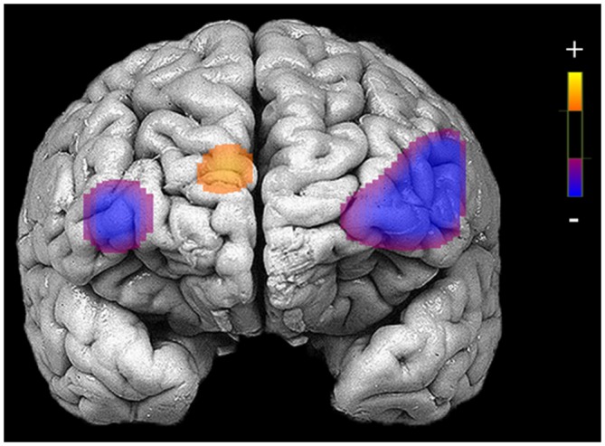
Garrison and colleagues recruited experienced meditators and non-meditators and scanned their brains (functional Magnetic Imaging, fMRI) while they either followed meditation instructions or an active mental task, making decisions as to whether adjectives applied to the self or to case. As expected the meditators reported less mind wandering during meditation. Importantly, they found that the meditators had significantly lower activity in the Default Mode Network (DMN) than the controls during the meditations but not during the active task.


These findings are important in that they demonstrate that the reduction in the DMN activity is not due to just any active mental task but specifically to meditation. The results also replicate the finding that meditation lowers mind wandering and the activity of the DMN. Hence meditation in particular appears to have the ability to reduce mind wandering, improving focus, by decreasing the activity of the brain system responsible for mind wandering.


So, reduce brain induced mind wandering with meditation.


“new knowledge about the default mode network and the self-reflecting thoughts that it stimulates may facilitate our understanding of how we function in our daily lives. We are more than intellect or the motor control of arms and legs, which is often the focus of brain researchers. Perhaps it may at times be good to know that our brain actually gives us room for our spontaneous thoughts and the associations and emotions that may at first seem a bit weird.” – Marcus Raichle


CMCS – Center for Mindfulness and Contemplative Studies


This and other Contemplative Studies posts are also available on Google+ https://plus.google.com/106784388191201299496/posts


Study Summary

Garrison, K. A., Zeffiro, T. A., Scheinost, D., Constable, R. T., & Brewer, J. A. (2015). Meditation leads to reduced default mode network activity beyond an active task. Cognitive, Affective & Behavioral Neuroscience, 15(3), 712–720. http://doi.org/10.3758/s13415-015-0358-3


Abstract

Meditation has been associated with relatively reduced activity in the default mode network, a brain network implicated in self-related thinking and mind wandering. However, previous imaging studies have typically compared meditation to rest despite other studies reporting differences in brain activation patterns between meditators and controls at rest. Moreover, rest is associated with a range of brain activation patterns across individuals that has only recently begun to be better characterized. Therefore, this study compared meditation to another active cognitive task, both to replicate findings that meditation is associated with relatively reduced default mode network activity, and to extend these findings by testing whether default mode activity was reduced during meditation beyond the typical reductions observed during effortful tasks. In addition, prior studies have used small groups, whereas the current study tested these hypotheses in a larger group. Results indicate that meditation is associated with reduced activations in the default mode network relative to an active task in meditators compared to controls. Regions of the default mode showing a group by task interaction include the posterior cingulate/precuneus and anterior cingulate cortex. These findings replicate and extend prior work indicating that suppression of default mode processing may represent a central neural process in long-term meditation, and suggest that meditation leads to relatively reduced default mode processing beyond that observed during another active cognitive task.

https://www.ncbi.nlm.nih.gov/pmc/articles/PMC4529365/


Improve Brain Processing of Emotions in the Elderly with Meditation


By John M. de Castro, Ph.D.


“Yet until recently little was known about how a few hours of quiet reflection each week could lead to such an intriguing range of mental and physical effects. Now, as the popularity of mindfulness grows, brain imaging techniques are revealing that this ancient practice can profoundly change the way different regions of the brain communicate with each other – and therefore how we think – permanently.” –  Tom Ireland


Meditation training has been shown to alter the nervous system, increasing the size and connectivity of structures associated with present moment awareness, higher level thinking, and regulation of emotions, while decreasing the size and connectivity of structures associated with mind wandering and self-referential thinking, known as the Default Mode Network (DMN). The brain is capable of changing and adapting in a process called neuroplasticity. As a result, the neural changes produced by meditation training become relatively permanent.


Meditation training has also been shown to produce improvements in emotion regulation. Most of the research to date on the neural systems altered by meditation training has focused on higher level cortical centers. But, emotions involve lower centers such as those located in the Pons in primitive brain stem. In today’s Research News article “Pons to Posterior Cingulate Functional Projections Predict Affective Processing Changes in the Elderly Following Eight Weeks of Meditation Training.” See:

https://www.facebook.com/ContemplativeStudiesCenter/photos/a.628903887133541.1073741828.627681673922429/1349231245100798/?type=3&theater

or see summary below or view the full text of the study at:

http://www.ncbi.nlm.nih.gov/pmc/articles/PMC5006446/

Shao and colleagues investigate the changes in connectivity between the Pons sites involved in emotions and the Posterior Cingulate Cortex and the Precuneus area which are important in the Default Mode Network (DMN). They recruited elderly (>60 years of age) participants with no experience with meditation or relaxation training through newspaper ads. They were randomly assigned to receive 8-weeks of either meditation training or relaxation training. Training occurred with 1.5-hour training sessions occurring 3 time per week. All participants received functional Magnetic Resonance Imaging (fMRI) brain scans and were tested with an emotion processing task both before and after training.


They found that after meditation training the elderly participants had moderated emotional responses that converged toward the middle, such that positive emotions were not as positive and arousing while negative emotions were not as negative and arousing as prior to training. This effect did not occur in the relaxation trained participants. They also found increased functional connectivity between the Pons emotion centers and the Posterior Cingulate Cortex and the Precuneus area components of the Default Mode Network (DMN). The increased connectivity was primarily in the Pons to DMN direction. In addition, the greater the change in the connectivity the greater the reduction in negative emotional responses by the participants. Again, these effect did not occur in the relaxation trained participants.


These are interesting results that extend the previous findings on improved emotion regulation after meditation training as the training was found to moderate emotional reactivity, making both positive and negative emotions less extreme. They further showed that this moderation of emotions is associated with increased connectivity between the areas of primitive emotion in the Pontine brain stem and the higher level Default Mode Network (DMN) procession in the Cerebral Cortex. These effects were shown to be due to the meditation training as relaxation training did not produce them.


One of the ways that emotions can get out of hand is by ruminating about past emotional responses and worrying about future emotional responses. This can increase the magnitude of emotional responses. Rumination and worry is the role of the DMN. The present research suggests that the improve emotion regulation seen after meditation training may be due to the increased influence of lower emotion centers on reducing the activation of the brain areas responsible for rumination and worry. In that way emotions can be experienced and analyzed as real time experiences and not amplified beyond their actual magnitude. This is a tremendous benefit of meditation training, allowing for more realistic appraisal of emotions.


So, improve brain processing of emotions in the elderly with meditation.


“One way to do this is mindfulness meditation, in which you observe your thoughts and feelings with the objectivity of a disinterested, nonjudgmental witness. This form of mental training gives you “the wherewithal to pause, observe how easily the mind can exaggerate the severity of a setback, note that it as an interesting mental process, and resist getting drawn into the abyss,” – Richie Davidson


CMCS – Center for Mindfulness and Contemplative Studies


This and other Contemplative Studies posts are also available on Google+ https://plus.google.com/106784388191201299496/posts


Study Summary

Robin Shao, Kati Keuper, Xiujuan Geng, Tatia M.C. Lee. Pons to Posterior Cingulate Functional Projections Predict Affective Processing Changes in the Elderly Following Eight Weeks of Meditation Training. EBioMedicine. 2016 Aug; 10: 236–248. Published online 2016 Jun 15. doi: 10.1016/j.ebiom.2016.06.018


Abstract

Evidence indicates meditation facilitates affective regulation and reduces negative affect. It also influences resting-state functional connectivity between affective networks and the posterior cingulate (PCC)/precuneus, regions critically implicated in self-referential processing. However, no longitudinal study employing active control group has examined the effect of meditation training on affective processing, PCC/precuneus connectivity, and their association. Here, we report that eight-week meditation, but not relaxation, training ‘neutralized’ affective processing of positive and negative stimuli in healthy elderly participants. Additionally, meditation versus relaxation training increased the positive connectivity between the PCC/precuneus and the pons, the direction of which was largely directed from the pons to the PCC/precuneus, as revealed by dynamic causal modeling. Further, changes in connectivity between the PCC/precuneus and pons predicted changes in affective processing after meditation training. These findings indicate meditation promotes self-referential affective regulation based on increased regulatory influence of the pons on PCC/precuneus, which new affective-processing strategy is employed across both resting state and when evaluating affective stimuli. Such insights have clinical implications on interventions on elderly individuals with affective disorders.

http://www.ncbi.nlm.nih.gov/pmc/articles/PMC5006446/


Improve the Brain’s Capacity for Creative Problem Solving with Meditation

By John M. de Castro, Ph.D.


“meditation is the best approach we know of allowing the mind to relax and have the ability to make a surge of insight wisdom. One does not power through an “Aha! Moment” with strain and determination. Thinking and more thinking does not generate that moment of brilliance that we are pursuing. The quick flash of energy is actually the subconscious mind surging a new perspective to the conscious mind. The conscious mind is more likely to recognize the gift of brilliance that the subconscious mind has sent it, if the conscious mind is in a calm state.” – Lindsay Leimbach


The problem solving ability of humans has been a key to their dominance of their environment. So, it’s important that we understand it and discover how to train it and maximize it. Problem solving most frequently involves logic and reasoning, sometimes along with mathematics. In this case focused attention is the key. The mind wandering off topic interferes with the concentration required for obtaining the solution. But, when a solution does not occur and the individual fails to solve the problem a completely different process transpires producing insight. If logic and reason fail, then fanciful and out-of-the box thinking may be needed. In this case mind wandering, taking the thought process away from the failed logical strategy, is superior, often producing a solution in a flash, an “aha” moment. In this case focused attention prevents the individual from seeing an unusual or creative solution. While the mind wandering off topic increases the discursive thinking that is required for obtaining the insightful solution.


Mindfulness is the ability to focus on what is transpiring in the present moment. It involves a greater emphasis on attention to the immediate stimulus environment. Mindful people generally have better attentional abilities and have fewer intrusive thoughts and less mind wandering. As a result, mindfulness has been shown to be associated with differences in thought processes. Most of the time these differences are associated with beneficial results, but sometimes they can lead to negative outcomes including a greater tendency to have false memories. So mindfulness should improve problem solving involving logic, reason, and focused attention, while it should interfere with insightful, creative problem solving.


These two forms of problem solving are, in general, associated with different neural systems. Focused attention involves a number of brain structures centered in the frontal lobes. Creative, discursive thinking involves a system of structures known as the Default Mode Network (DMN) involving the parietal lobe, cingulate cortex, and insula. One way to investigate the influence of mindfulness on creative problem solving is to look at the activity of these two systems during problem solving and insight before and after mindfulness training.


In today’s Research News article “Short-term meditation modulates brain activity of insight evoked with solution cue.” See:

https://www.facebook.com/ContemplativeStudiesCenter/photos/a.628903887133541.1073741828.627681673922429/1341430935880829/?type=3&theater

or see summary below or view the full text of the study at:

http://www.ncbi.nlm.nih.gov/pmc/articles/PMC4994853/

Ding and colleagues randomly assigned participants to receive either meditation training, integrative body–mind training (IBMT), or simple relaxation for 10 days, 30 minutes per day. Before and after the training they had the participants perform a task that requires creative thought known as the Remote Associations Test, which also frequently produces “aha’ moments when the solution is seen. The task was performed while the participants’ neural activity was measured in a functional Magnetic Resonance (f-MRI) scanner.


They found that after the meditation training the participants were significantly better at the Remote Associations Test. They then looked at the brain activity when the solution was not found and they were presented with the solution, producing an “aha’ moment. They found that after meditation training the brain activity of the two groups differed in this “aha’ moment. There was significantly greater activation found in the right cingulate gyrus, insula, putamen, as well as in the right inferior frontal gyrus, and the bilateral middle frontal gyrus, inferior parietal lobule and superior temporal gyrus. Some of these structures are involved in focused logical reasoning while others are part of the mind wandering system (DMN).


The “aha’ moment produced in this study involves logic in analyzing the possibilities and seeing that the proposed solution actually solves the problem and also the discursive, creative though to see how the unusual (remote) solution is appropriate. These findings suggest that mindfulness training alters the nervous system making it able to both utilize the focused logical systems and the mind wandering, discursive systems better to solve this kind of problem. Hence, it appears that meditation training enhances both logical and insightful problem solving by enhancing the activity of their underlying neural systems.


So, improve the brain’s capacity for creative problem solving with meditation.


“If, individually and collectively, we can be more mindful of the way we employ problem-solving strategies, we could release a great deal of human potential from the damaging effects of stress.” – Matthew Kalman Mezey


CMCS – Center for Mindfulness and Contemplative Studies


This and other Contemplative Studies posts are also available on Google+ https://plus.google.com/106784388191201299496/posts


Study Summary

Ding, X., Tang, Y.-Y., Cao, C., Deng, Y., Wang, Y., Xin, X., & Posner, M. I. (2015). Short-term meditation modulates brain activity of insight evoked with solution cue. Social Cognitive and Affective Neuroscience, 10(1), 43–49. http://doi.org/10.1093/scan/nsu032


Abstract

Meditation has been shown to improve creativity in some situation. However, little is known about the brain systems underling insight into a problem when the person fails to solve the problem. Here, we examined the neural correlation using Chinese Remote Association Test, as a measure of creativity. We provide a solution following the failure of the participant to provide one. We examine how meditation in comparison with relaxation influences the reaction of the participant to a correct solution. The event-related functional magnetic resonance imaging showed greater activity, mainly distributed in the right cingulate gyrus (CG), insula, putamen, inferior frontal gyrus (IFG), and the bilateral middle frontal gyrus (MFG), the inferior parietal lobule (IPL) and the superior temporal gyrus (STG). This pattern of activation was greater following 5 h of meditation training than the same amount of relaxation. Based on prior research, we speculate on the function of this pattern of brain activity: (i) CG may be involved in detecting conflict and breaking mental set, (ii) MFG/IFG may play an important role in restructuring of the problem representation, (iii) insula, IPL and STG may be associated with error detection, problem understanding or general attentive control and (iv) putamen may be activated by ‘Aha’ feeling.

http://www.ncbi.nlm.nih.gov/pmc/articles/PMC4994853/


Reduce Brain Asymmetries with Meditation

By John M. de Castro, Ph.D.


“The practice appears to have an amazing variety of neurological benefits – from changes in grey matter volume to reduced activity in the “me” centers of the brain to enhanced connectivity between brain regions.” – Alice G. Walton


The higher centers of the human brain are contained in two separate hemispheres. Over evolutionary history these hemispheres have taken on different functions. For most people the left hemisphere has become responsible for language, mathematics, logic, and the activities of the right side of the body while the right hemisphere has become responsible for spatial ability, emotions, artistic expression, and the activities of the left side of the body. These differences in function are accompanied with differences in the sizes of the structures and their interconnectivity on the two sides.


Contemplative practices such as meditation, yoga, and tai chi have been shown to produce changes in the sizes, activity, and connectivity of brain structures through a process called neuroplasticity. The degree to which these practices might alter the differences between the hemispheres, however, has not been extensively studied. In today’s Research News article “Shifting brain asymmetry: the link between meditation and structural lateralization.” See:

https://www.facebook.com/ContemplativeStudiesCenter/photos/a.628903887133541.1073741828.627681673922429/1340603669296889/?type=3&theater

or see summary below or view the full text of the study at:

http://www.ncbi.nlm.nih.gov/pmc/articles/PMC4994843/

Kurth and colleagues investigate the effects of long-term meditation practices on the differences between the hemispheres. They recruited adult long-term meditators (> 4 years, mean 19 years of practice) and matched control subjects and subjected their brains to Magnetic Resonance Imaging (MRI) scans. These brain images were then analyzed for structural differences between the hemispheres.


They found that the meditators had larger parietal lobes and less difference between the two hemispheres than the non-meditators who had right hemispheres larger than the left. This difference was particularly centered in a parietal lobe structure called the precuneus. In addition, the greater the number of years of meditation practice, the larger the left hemisphere and the smaller the right hemisphere producing less of a difference between the sides. This decrease in the differences between the hemispheres was also present in the anterior cingulate cortex with the greater the number of practice years the smaller the differences.


These are interesting results that suggest that meditation practice reduces the differences between the hemispheres in the parietal lobe and anterior cingulate cortex. These areas are components of a neural system termed the Default Mode Network (DMN) which is activated during unfocused conscious activity deemed mind wandering. Meditation, with its emphasis on focused attention has been shown to reduce the size and connectivity of the DMN and mind wandering. Kurth and colleagues’ study suggests that meditation also reduces the differences in the sizes of the DMN in the two hemispheres.


These interesting findings suggest that long-term meditation practice may produce greater balance between the two hemispheres and this suggests greater intermixing of their separated functions; integrating the tow hemispheres’ functions. So, logical thinking and emotions intermix, art and language work together, the two sides of the body work more harmoniously and the individual attains a greater integrated wholeness. This is certainly one of the goals of meditation practice and the change in the hemispheric differences may signal success.


So, reduce brain asymmetries with meditation.


“The impact that mindfulness exerts on our brain is borne from routine: a slow, steady, and consistent reckoning of our realities, and the ability to take a step back, become more aware, more accepting, less judgmental, and less reactive. Just as playing the piano over and over again over time strengthens and supports brain networks involved with playing music, mindfulness over time can make the brain, and thus, us, more efficient regulators, with a penchant for pausing to respond to our worlds instead of mindlessly reacting.” –  Jennifer Wolkin


CMCS – Center for Mindfulness and Contemplative Studies


This and other Contemplative Studies posts are also available on Google+ https://plus.google.com/106784388191201299496/posts


Study Summary

Kurth, F., MacKenzie-Graham, A., Toga, A. W., & Luders, E. (2015). Shifting brain asymmetry: the link between meditation and structural lateralization. Social Cognitive and Affective Neuroscience, 10(1), 55–61. http://doi.org/10.1093/scan/nsu029


Abstract

Previous studies have revealed an increased fractional anisotropy and greater thickness in the anterior parts of the corpus callosum in meditation practitioners compared with control subjects. Altered callosal features may be associated with an altered inter-hemispheric integration and the degree of brain asymmetry may also be shifted in meditation practitioners. Therefore, we investigated differences in gray matter asymmetry as well as correlations between gray matter asymmetry and years of meditation practice in 50 long-term meditators and 50 controls. We detected a decreased rightward asymmetry in the precuneus in meditators compared with controls. In addition, we observed that a stronger leftward asymmetry near the posterior intraparietal sulcus was positively associated with the number of meditation practice years. In a further exploratory analysis, we observed that a stronger rightward asymmetry in the pregenual cingulate cortex was negatively associated with the number of practice years. The group difference within the precuneus, as well as the positive correlations with meditation years in the pregenual cingulate cortex, suggests an adaptation of the default mode network in meditators. The positive correlation between meditation practice years and asymmetry near the posterior intraparietal sulcus may suggest that meditation is accompanied by changes in attention processing.

http://www.ncbi.nlm.nih.gov/pmc/articles/PMC4994843/


Permanently Improve Brain Dynamics with Meditation Practice

By John M. de Castro, Ph.D.


“The nature of brain vs. mind vs. consciousness is a question that may remain unanswerable but the important thing to remember is to live in awareness of how interconnected each of these aspects of yourself really are. Using these tips, you will grow in self-awareness and strengthen your connections with the deepest parts of your consciousness.” – Operation-Meditation


We spend a tremendous amount of our time with our minds wandering and not on the task or the environment at hand. We daydream, plan for the future, review the past, ruminate on our failures, exalt in our successes. In fact, we spend almost half of our waking hours off task with our mind wandering. You’d think that if we spend so much time doing this it must be enjoyable. But, in fact research has shown that when our mind is wandering we are actually unhappy compared to when we are paying attention to what is at hand.


A system of the brain known as the Default Mode Network (DMN) becomes active during wind wandering and relatively quiet during focused on task behavior. It is involved when we are engaged in internally focused tasks such as recalling deeply personal memories, daydreaming, sleeping, imagining the future and trying to take the perspective of others. The DMN involves neural structures including the medial prefrontal cortex, anterior and posterior cingulate cortices, precuneus, inferior parietal cortex, and lateral temporal cortex. These areas of the DMN are functionally connected, such that they are simultaneously active during mind wandering.


Meditation is known to reduce the size and activity of the Default Mode Network (DMN) through a process known as neuroplasticity where the size and connectivity of neural structures are modified by experience. In today’s Research News article “Temporal Dynamics of the Default Mode Network Characterize Meditation-Induced Alterations in Consciousness.” See:

https://www.facebook.com/ContemplativeStudiesCenter/photos/a.628903887133541.1073741828.627681673922429/1330785286945394/?type=3&theater

or see summary below or view the full text of the study at:

http://www.ncbi.nlm.nih.gov/pmc/articles/PMC4956663/

Panda and colleagues further investigate the effects of meditation on the DMN. They recruited male experienced meditators with over 20 years of daily meditation experience and a matched group of non-meditators. They then simultaneously measured brain activity with both functional Magnetic Resonance Imaging (f-MRI) and with electroencephalography (EEG) both while the participants were at rest and during meditation.


They found that the meditators in comparison to the non-meditators had reduced connectivity of the posterior cingulate cortex that was further reduced during meditation. The posterior cingulate cortex is a key structure of the DMN. This suggests that meditation reduces the ability of the DMN to produce mind wandering. They also found increased connectivity in the middle frontal and middle temporal gyri that increased further during meditation. These are both structures associated with focused attention. These effects suggest that meditation experience increases the activity of the attentional system while decreasing the activity of the mind wandering system.


Panda and colleagues analyzed the electroencephalography (EEG) signals recorded simultaneously with the functional Magnetic Resonance Imaging (f-MRI) characterizing periods of stable activity over brief periods of time called microstates. They found that meditators had these stable states more often and for longer periods of time both at rest and during meditation. The more years of meditation practice the greater the increased stability in the DMN. With the non-meditators these stable states in the DMN occurred more frequently and for longer periods during meditation. These findings suggest that meditation increases stability of the DMN and extensive meditation experience permanently alters the DMN to be more stable all of the time.


These findings suggest that meditation reduced the activity of the Default Mode Network (DMN) briefly during meditation or permanently with experience with meditation. This further suggests that meditation experience permanently alters the tendency for the mind to wander both during meditation and also at rest. The studies of the alterations of the nervous system with meditation suggest that the changes in the states of consciousness seen with meditation are reflected in changes in the underlying neural systems, producing great focus and less mind wandering.


So, permanently improve brain dynamics with meditation practice.


“we do know that meditation is a tool that can change brain activity to bring it closer to this optimal, high-performance brain wave pattern. Subjective experiences of meditators with this optimal brain wave pattern reveal that they experience bliss in this state.” – Shanida Nataraja


CMCS – Center for Mindfulness and Contemplative Studies


This and other Contemplative Studies posts are also available on Google+ https://plus.google.com/106784388191201299496/posts


Study Summary

Panda, R., Bharath, R. D., Upadhyay, N., Mangalore, S., Chennu, S., & Rao, S. L. (2016). Temporal Dynamics of the Default Mode Network Characterize Meditation-Induced Alterations in Consciousness. Frontiers in Human Neuroscience, 10, 372. http://doi.org/10.3389/fnhum.2016.00372

Abstract

Current research suggests that human consciousness is associated with complex, synchronous interactions between multiple cortical networks. In particular, the default mode network (DMN) of the resting brain is thought to be altered by changes in consciousness, including the meditative state. However, it remains unclear how meditation alters the fast and ever-changing dynamics of brain activity within this network. Here we addressed this question using simultaneous electroencephalography (EEG) and functional magnetic resonance imaging (fMRI) to compare the spatial extents and temporal dynamics of the DMN during rest and meditation. Using fMRI, we identified key reductions in the posterior cingulate hub of the DMN, along with increases in right frontal and left temporal areas, in experienced meditators during rest and during meditation, in comparison to healthy controls (HCs). We employed the simultaneously recorded EEG data to identify the topographical microstate corresponding to activation of the DMN. Analysis of the temporal dynamics of this microstate revealed that the average duration and frequency of occurrence of DMN microstate was higher in meditators compared to HCs. Both these temporal parameters increased during meditation, reflecting the state effect of meditation. In particular, we found that the alteration in the duration of the DMN microstate when meditators entered the meditative state correlated negatively with their years of meditation experience. This reflected a trait effect of meditation, highlighting its role in producing durable changes in temporal dynamics of the DMN. Taken together, these findings shed new light on short and long-term consequences of meditation practice on this key brain network.

http://www.ncbi.nlm.nih.gov/pmc/articles/PMC4956663/

Reduce Mind Wandering Produced by the Brain with Mindfulness


By John M. de Castro, Ph.D.


“the default mode network always takes you somewhere other than where you are. You are not focused on the immediate environment and will also be “time-travelling” in the past or future – never in the present moment. . . Interestingly, researchers have found that the default mode network is less active when people meditate.” – Mindful Call


Mindfulness is the ability to focus on what is transpiring in the present moment. It involves a greater emphasis on attention to the immediate stimulus environment. Mindful people generally have better attentional abilities and have fewer intrusive thoughts and less mind wandering. As a result, mindfulness has been shown to be associated with differences in thought processes. A system of the brain known as the Default Mode Network (DMN) becomes active during mind wandering and relatively quiet during focused on task behavior. It is involved when we are engaged in internally focused tasks such as recalling deeply personal memories, daydreaming, sleeping, imagining the future and trying to take the perspective of others. The DMN involves neural structures including the medial prefrontal cortex, anterior and posterior cingulate cortices, precuneus, inferior parietal cortex, and lateral temporal cortex. These areas of the DMN are functionally connected, such that they are simultaneously active during mind wandering.


Mindfulness training has been shown to alter the size and activity of neural structures including reducing the size and activity in the Default Mode Network (DMN) as a result of neuroplasticity. The DMN, however, is a network of interconnected structures which are, in turn, connected to a number of other neural structures. This interconnectivity reflects the ability of these structures to affect other structures in the brain, in other words, the effects of mind wandering on other brain systems. Hence, it is important to investigate the effects of mindfulness training on the functional connectivity of the structures of the Default Mode Network (DMN) and the neural structures connected to these structures.


In today’s Research News article “Data for default network reduced functional connectivity in meditators, negatively correlated with meditation expertise.” See:

https://www.facebook.com/ContemplativeStudiesCenter/photos/a.628903887133541.1073741828.627681673922429/1328281653862424/?type=3&theater

or see summary below or view the full text of the study at:

http://www.sciencedirect.com/science/article/pii/S2352340916304504

Berkovich-Ohana and colleagues performed functional Magnetic Resonance Imaging (fMRI) of the brains of experienced meditators in comparison to similar individuals who did not meditate while at rest and during meditation.


They found that at rest the functional connectivity within the structures of the DMN and within the neural structures associated with vision were lower in meditators and that this functional connectivity decreased during meditation. They also found that the meditators had greater functional connectivity between the DMN and the visual system structures than the non-meditators and this also decreased during meditation. In addition, they found that the greater the experience with meditation the lower the functional connectivity between the DMN and other neural structures. These findings suggest that meditation lowers the ability of the structures associated with mind wandering to affect other structures in the nervous system. They also suggest that meditation practice produces less internal connectivity within these structures underlying mind wandering. Finally, these findings suggest that visualizations dissociated from the focus of attention may be higher in the meditators. This may indicate that when meditators’ minds wander they contain more vivid visual imagery.


These data are interesting and demonstrate that meditation alters the internal connectivity of the structures that produce mind wandering and their ability to affect other neural structures. Hence, the functional connectivity of brain structures reflects the experiences of meditators of reduced mind wandering. Meditation appears to change the brain to produce less mind wandering.


So, reduce mind wandering produced by the brain with mindfulness.


“What may happen when people practise mindfulness is that, over time, this weakens the connection between their thalamus and the rest of the default mode network. Their trait mindfulness score would climb as a result and they might just become more mentally robust and less prone to depression and other mental illnesses. That really would be brain plasticity in action.” – Plastic Brain


CMCS – Center for Mindfulness and Contemplative Studies


This and other Contemplative Studies posts are also available on Google+ https://plus.google.com/106784388191201299496/posts


Study Summary

Berkovich-Ohana A, Harel M, Hahamy A, Arieli A, Malach R. Data for default network reduced functional connectivity in meditators, negatively correlated with meditation expertise. Data Brief. 2016 Jul 15;8:910-4. doi: 10.1016/j.dib.2016.07.015. eCollection 2016 Sep. PMID: 2750824


Abstract

FMRI data described here was recorded during resting-state in Mindfulness Meditators (MM) and control participants (see “Task-induced activity and resting-state fluctuations undergo similar alterations in visual and DMN areas of long-term meditators” Berkovich-Ohana et al. (2016) [1] for details). MM participants were also scanned during meditation. Analyses focused on functional connectivity within and between the default mode network (DMN) and visual network (Vis). Here we show data demonstrating that: 1) Functional connectivity within the DMN and the Visual networks were higher in the control group than in the meditators; 2) Data show an increase for the functional connectivity between the DMN and the Visual networks in the meditators compared to controls; 3) Data demonstrate that functional connectivity both within and between networks reduces during meditation, compared to the resting-state; and 4) A significant negative correlation was found between DMN functional connectivity and meditation expertise.

http://www.sciencedirect.com/science/article/pii/S2352340916304504

http://ac.els-cdn.com/S2352340916304504/1-s2.0-S2352340916304504-main.pdf?_tid=3b020506-60c4-11e6-bbcf-00000aacb360&acdnat=1471030840_5c9ab5e6d389861ca100e4384dec9a9f


Improve the Brain’s Emotional Responses with Mindfulness

By John M. de Castro, Ph.D.


“Meditation gives you the wherewithal to pause, observe how easily the mind can exaggerate the severity of a setback, and resist getting drawn back into the abyss.”— Richie Davidson


We’re very emotional creatures. Without emotion, life is flat and uninteresting. Emotions provide the spice of life. We are constantly having or reacting to emotions. We often go to great lengths in an attempt to create or keep positive emotions and conversely to avoid, mitigate, or get rid of negative emotions. They are so important to us that they affect mostly everything that we do and say and can even be determinants of life or death. Anger, fear, and hate can lead to murderous consequences. Anxiety and depression can lead to suicide. At the same time love, joy, and happiness can make life worth living. Our emotions also affect us physically with positive emotions associated with health, well-being, and longevity and negative emotions associated with stress, disease, and shorter life spans. The importance of emotions is only surpassed by our ignorance of them.


Emotions occur automatically and reflexively to particularly stimuli in the environment. For example, the sight of a snake almost universally evokes a fear response, or conversely the sight of a baby smiling almost universally evokes loving feelings. Psychologists have demonstrated that these reflexive emotional reactions can be transferred to other stimuli. This occurs in a process first described by Pavlov called classical conditioning. For example, seeing a snake in a flower pot can later produce fear responses to the flower pot itself. This is called emotional learning.


One of the most important effects of mindfulness training is improving emotion regulation. Research has demonstrated that people either spontaneously high in mindfulness or trained in mindfulness are better able to be completely in touch with their emotions and feel them completely, while being able to respond to them more appropriately and adaptively. In other words, mindful people are better able to experience yet control emotions. The influence of mindfulness training on emotional learning has not, however, been extensively studied.


In today’s Research News article “Mindfulness-Based Stress Reduction, Fear Conditioning, and The Uncinate Fasciculus: A Pilot Study.” See:

https://www.facebook.com/ContemplativeStudiesCenter/photos/a.628903887133541.1073741828.627681673922429/1312616582095598/?type=3&theater

or see summary below or view the full text of the study at:

http://www.ncbi.nlm.nih.gov/pmc/articles/PMC4908122/

Hölzel and colleagues randomly assigned adult participants to either receive an 8-week Mindfulness-Based Stress Reduction (MBSR) program or be placed on a wait-list. Prior to and after the 8-week training period, both groups were assessed for mindfulness, perceived stress, and emotion regulation ability. They were also tested with a 2-day fear conditioning emotional learning, extinction and retention of extinction procedure which occurred while the participants laid in a Magnetic Resonance Imaging (MRI) scanner. The conditioning occurred to red and blue lights paired with a mild electric shock. Then only one light and not the other was paired with shock so that the fear response to one would decline in strength (extinguish).


They found that the MBSR training produced significant improvements in mindfulness, emotion regulation and perceived stress. In addition, the more home practice the participants engaged in the greater the improvement. They found that the MBSR participants retained the fear conditioning from prior to, to after the conditioning while the control group did not. In addition, MBSR was found to produce a significant increase in the fiber density, axonal diameter, and myelination of the Uncinate fasciclus; in other words, it increased the size of this fiber tract that interconnects the limbic system to the frontal lobes. Aslo, the greater the increase in the size of the Uncinate, the stronger the increase in the strength of the fear conditioning.


It has been repeatedly demonstrated that mindfulness (MBSR) training increases emotion regulation and decreases stress responses. So, these results in the present study were no surprise. The increased retention of the fear conditioning found after mindfulness training is interesting and suggests that the training did what is was supposed to, increasing attentiveness to environmental stimuli and thus making the individual more responsive to them over longer periods. It is possible that mindfulness training, by improving emotion regulation and stress responding allowed for better appreciation and control of prior emotional conditioning. Hence, mindfulness training appears to make us better at being attentive to and regulating both our primary and secondary (learned) emotions.


The improved retention of the fear conditioning may also result from the increased size of the Uncinate fasciculus which connects the limbic system which is known to be involved in emotions to the frontal lobes which are known to be involved in emotion regulation. Hence, the MBSR training appears to have altered the brain to accentuate the processing and regulation of emotional signals. These kind of changes in brain structure, known as neuroplasticity, are commonly found after mindfulness training. In the case of the present study the change in the brain produced by mindfulness training appears to alter the individual’s responsiveness and control of their emotions.


So, improve the brain’s emotional responses with mindfulness.


“Mindfulness practice isn’t meant to eliminate thinking but aims rather to help us know what we’re thinking when we’re thinking it, just as we want to know what we’re feeling when we’re feeling it. Mindfulness allows us to watch our thoughts, see how one thought leads to the next, decide if we’re heading toward an unhealthy path, and if so, let go and change directions. It allows us to see that who we are is much more than a fearful or envious or angry thought. We can rest in the awareness of the thought, in the compassion we extend to ourselves if the thought makes us uncomfortable, and in the balance and good sense we summon as we decide whether and how to act on the thought.” – Sharon Salzberg


CMCS – Center for Mindfulness and Contemplative Studies


This and other Contemplative Studies posts are also available on Google+ https://plus.google.com/106784388191201299496/posts


Study Summary

Hölzel, B. K., Brunsch, V., Gard, T., Greve, D. N., Koch, K., Sorg, C., … Milad, M. R. (2016). Mindfulness-Based Stress Reduction, Fear Conditioning, and The Uncinate Fasciculus: A Pilot Study. Frontiers in Behavioral Neuroscience, 10, 124. http://doi.org/10.3389/fnbeh.2016.00124


Abstract

Mindfulness has been suggested to impact emotional learning, but research on these processes is scarce. The classical fear conditioning/extinction/extinction retention paradigm is a well-known method for assessing emotional learning. The present study tested the impact of mindfulness training on fear conditioning and extinction memory and further investigated whether changes in white matter fiber tracts might support such changes. The uncinate fasciculus (UNC) was of particular interest in the context of emotional learning. In this pilot study, 46 healthy participants were quasi-randomized to a Mindfulness-Based Stress Reduction (MBSR,N = 23) or waitlist control (N = 23) group and underwent a two-day fear conditioning, extinction learning, and extinction memory protocol before and after the course or control period. Skin conductance response (SCR) data served to measure the physiological response during conditioning and extinction memory phases. Diffusion tensor imaging (DTI) data were analyzed with probabilistic tractography and analyzed for changes of fractional anisotropy in the UNC. During conditioning, participants were able to maintain a differential response to conditioned vs. not conditioned stimuli following the MBSR course (i.e., higher sensitivity to the conditioned stimuli), while controls dropped the response. Extinction memory results were not interpretable due to baseline differences. MBSR participants showed a significant increase in fractional anisotropy in the UNC, while controls did not (group by time interaction missed significance). Pre-post changes in UNC were correlated with changes in the response to the conditioned stimuli. The findings suggest effects of mindfulness practice on the maintenance of sensitivity of emotional responses and suggest underlying neural plasticity.

http://www.ncbi.nlm.nih.gov/pmc/articles/PMC4908122/


Change your Brain for the Better with Mindfulness Based Stress Reduction

By John M. de Castro, Ph.D.


“Neuroscientists have also shown that practicing mindfulness affects brain areas related to perception, body awareness, pain tolerance, emotion regulation, introspection, complex thinking, and sense of self. . . When we take a seat, take a breath, and commit to being mindful, particularly when we gather with others who are doing the same, we have the potential to be changed.” – Christina Congleton


The nervous system is a dynamic entity, constantly changing and adapting to the environment. It will change size, activity, and connectivity in response to experience. For example, the brain area that controls the right index finger has been found to be larger in blind subjects who use braille than in sighted individuals.  Similarly, cab drivers in London who navigate the twisting streets of the city, have a larger hippocampus, which is involved in spatial navigation, than predefined route bus drivers. These changes in the brain are called neuroplasticity. Over the last decade neuroscience has been studying the effects of contemplative practices on the brain and has identified neuroplastic changes in widespread areas. In other words, meditation practice appears to mold and change the brain, producing psychological, physical, and spiritual benefits.


The seemingly simple behavior of meditation is actually quite complex. Adding to the complexity is that there are a variety of different meditation techniques. To begin to understand exactly how meditation works to produce its benefit, it is important to determine what works best and what doesn’t. So, there is a need to test and compare the effects of a variety of techniques and variations. There has been some work investigating the neuroplastic changes resulting from a number of different types of meditation techniques. But more work is needed.


Mindfulness Based Stress Reduction (MBSR) is a practice widely used particularly to treat mental and physical conditions. It is, in fact, an amalgam of three mindfulness practice techniques; meditation, body scan, and yoga. It is not known if this combination of practices has the same effects on the nervous system as simple long-term meditation practice. In today’s Research News article “8-week Mindfulness Based Stress Reduction induces brain changes similar to traditional long-term meditation practice – A systematic review.” See:

https://www.facebook.com/ContemplativeStudiesCenter/photos/a.628903887133541.1073741828.627681673922429/1309864165704173/?type=3&theater

or see summary below. Gotink and colleagues review the published research literature on the effects of Mindfulness Based Stress Reduction (MBSR) training on the brain and compare it to the effects of long-term meditation. Participants in the studies were adults who were provided an 8-week MBSR program and had their brains scanned with functional Magnetic Resonance Imaging (f-MRI).


They found that the literature reported that 8 weeks of MBSR training produced changed activity and functional connectivity in the prefrontal cortex, hippocampus, insula, and cingulate cortex. These are all brain structures that are involved in emotion regulation and behavioral response inhibition and control. In addition, the studies report decreased activity and size of the amygdala. This is a structure involved in activation and emotionality. These are very similar to the neural changes that have been reported for long-term meditators. So, it would appear that and 8-week MBSR training is sufficient to produce major changes in the nervous system reflecting changes in the psychological and emotional aspects of the individual. The practitioner’s brain is changed in such a way as to make them better in charge of their emotions and behavior.


So, change your brain for the better with mindfulness based stress reduction.


“Noticing the differences between sense and story, between primary experience-dependent ‘bottom-up’ input and the secondary ‘top-down’ chatter of prior learning becomes a fundamental tool of the mindfulness approach. Once this distinction, this noticing of the contents of the mind, is readily accessible through intentional practice, the capacity to alter habitual patterns is created and the possibility becomes available for relief from self-preoccupied rumination, self-defeating thought-patterns, negative autobiographical narratives and maladaptive patterns of emotional reactivity.” – Daniel J. Siegel


CMCS – Center for Mindfulness and Contemplative Studies


This and other Contemplative Studies posts are also available on Google+ https://plus.google.com/106784388191201299496/posts


Study Summary

Gotink RA, Meijboom R, Vernooij MW, Smits M, Hunink MG. 8-week Mindfulness Based Stress Reduction induces brain changes similar to traditional long-term meditation practice – A systematic review. Brain Cogn. 2016 Jul 15;108:32-41. doi: 10.1016/j.bandc.2016.07.001. [Epub ahead of print]


Highlights

  • Literature reports that long-term meditators show altered brain activations and structure.
  • Post-MBSR, prefrontal cortex, insula, cingulate cortex and hippocampus show similar results to traditional meditation.
  • In addition, the amygdala shows earlier deactivation, less gray matter and better connectivity.
  • These changes indicate a neuronal working mechanism of MBSR.

Abstract: The objective of the current study was to systematically review the evidence of the effect of secular mindfulness techniques on function and structure of the brain. Based on areas known from traditional meditation neuroimaging results, we aimed to explore a neuronal explanation of the stress-reducing effects of the 8-week Mindfulness Based Stress Reduction (MBSR) and Mindfulness Based Cognitive Therapy (MBCT) program.

Methods: We assessed the effect of MBSR and MBCT (N = 11, all MBSR), components of the programs (N = 15), and dispositional mindfulness (N = 4) on brain function and/or structure as assessed by (functional) magnetic resonance imaging. 21 fMRI studies and seven MRI studies were included (two studies performed both).

Results: The prefrontal cortex, the cingulate cortex, the insula and the hippocampus showed increased activity, connectivity and volume in stressed, anxious and healthy participants. Additionally, the amygdala showed decreased functional activity, improved functional connectivity with the prefrontal cortex, and earlier deactivation after exposure to emotional stimuli.

Conclusion: Demonstrable functional and structural changes in the prefrontal cortex, cingulate cortex, insula and hippocampus are similar to changes described in studies on traditional meditation practice. In addition, MBSR led to changes in the amygdala consistent with improved emotion regulation. These findings indicate that MBSR-induced emotional and behavioral changes are related to functional and structural changes in the brain.

Alter the Brain for Better Pain Management with Meditation

meditation pain2 Bilevicius

By John M. de Castro, Ph.D.


“For some people with chronic pain, mindful meditation is an appealing pain management option because it has an unusual benefit; it is something that you personally control. Unlike pain medications or medical procedures, meditation is not done to you, it is something you can do for yourself.” – Stephanie Burke


Pain can be difficult to deal with, particularly if it’s persistent. But, even short-term pain, acute pain, is unpleasant. Pain, however, is an important signal that there is something wrong or that damage is occurring. This signals that some form of action is needed to mitigate the damage. This is an important signal that is ignored at the individual’s peril. So, in dealing with pain, it’s important that pain signals not be blocked or prevented. They need to be perceived. Nevertheless, it would be useful to find ways to lower the intensity of perceived pain and improve recovery from painful stimuli.


Pain signals are processed in the brain and the state of the brain can alter the perception of pain. Indeed, pain is affected by the mind. The perception of pain can be amplified by the emotional reactions to it and also by attempts to fight or counteract it. On the other hand, pain perception can also be reduced by mental states. Indeed, contemplative practices have been shown to reduce both chronic and acute pain. These changes are reflected in the underlying processing of the pain signals in the nervous system. This suggests that mindfulness training produces long-lasting alterations of the neural circuits underlying pain processing.


In today’s Research News article “Altered Neural Activity Associated with Mindfulness during Nociception: A Systematic Review of Functional MRI.” See:

https://www.facebook.com/ContemplativeStudiesCenter/photos/a.628903887133541.1073741828.627681673922429/1299302553427001/?type=3&theater

or see summary below or view the full text of the study at:

http://www.ncbi.nlm.nih.gov/pmc/articles/PMC4931491/

Bilevicius and colleagues review the published research literature on the neural circuits involved in mindfulness’ alterations of perceived pain. In these studies, it was routinely reported that mindfulness produced a reduction in the anticipation of pain and pain unpleasantness. There were mixed findings in regard to whether the intensity of pain was reduced or not. Regardless, mindfulness appears to reduce unpleasantness without blocking the actual perception of pain.


They then looked at the reported effects of mindfulness training on brain activity as measured with functional Magnetic Resonance Imagery (fMRI). The published studies reported consistently that mindfulness training increased the activity of two key areas in pain processing, the Insula and the Anterior Cingulate Cortex in response to pain signals. On the other hand, mindfulness training produced decreased activity in response to pain in the Lateral Prefrontal Cortex. The ACC and the Insula are involved in processing stimuli originating in the present moment and their increased activity suggests that the mindfulness training altered the neural circuits involved in present moment awareness of pain signals. The Lateral Prefrontal Cortex, on the other hand, is associated with the awareness, cognitive processing, of pain. This suggests that mindfulness training produces a reduction in the thinking about pain.


These results suggest that mindfulness training produces lasting changes to the nervous system, sometimes called neuroplasticity. These changes altered the usual processing of pain signals. Although, the pain signals occurring in the present moment are heightened, they have less of an impact upon awareness and cognitive appreciation of pain. Mindfulness training, then appears to produce long-lasting changes in the brain that allow pain signals to be processed but reduce the psychological responses to pain, making it less unpleasant. The individual then can respond adaptively to the pain but not suffer as much.


So, alter the brain for better pain management with meditation.


“Imaging studies show that mindfulness soothes the brain patterns underlying pain and, over time, these changes take root and alter the structure of the brain itself, so that patients no longer feel pain with the same intensity. Many say that they barely notice it at all.” – Danny Penman


CMCS – Center for Mindfulness and Contemplative Studies


This and other Contemplative Studies posts are also available on Google+ https://plus.google.com/106784388191201299496/posts


Study Summary

Bilevicius, E., Kolesar, T. A., & Kornelsen, J. (2016). Altered Neural Activity Associated with Mindfulness during Nociception: A Systematic Review of Functional MRI. Brain Sciences, 6(2), 14. http://doi.org/10.3390/brainsci6020014


Abstract

Objective: To assess the neural activity associated with mindfulness-based alterations of pain perception. Methods: The Cochrane Central, EMBASE, Ovid Medline, PsycINFO, Scopus, and Web of Science databases were searched on 2 February 2016. Titles, abstracts, and full-text articles were independently screened by two reviewers. Data were independently extracted from records that included topics of functional neuroimaging, pain, and mindfulness interventions. Results: The literature search produced 946 total records, of which five met the inclusion criteria. Records reported pain in terms of anticipation (n = 2), unpleasantness (n = 5), and intensity (n = 5), and how mindfulness conditions altered the neural activity during noxious stimulation accordingly. Conclusions: Although the studies were inconsistent in relating pain components to neural activity, in general, mindfulness was able to reduce pain anticipation and unpleasantness ratings, as well as alter the corresponding neural activity. The major neural underpinnings of mindfulness-based pain reduction consisted of altered activity in the anterior cingulate cortex, insula, and dorsolateral prefrontal cortex.

http://www.ncbi.nlm.nih.gov/pmc/articles/PMC4931491/


Reduce Self-harm with Mindfulness-based Dialectical Behavior Therapy

DBT self-harm2 Ruocco


By John M. de Castro, Ph.D.


“People who self-injure have often found themselves either the victims of abuse or otherwise powerless and helpless in the midst of terrible circumstances. Self-abuse often provides an escape from overwhelming feelings of isolation, fear, humiliation or fury. Some who self-injure explain that it gives a sense of control in a world where they feel helpless. It can also be a physical sign of emotional pain.” – CHRISTY MATTA


Self-injury is a disturbing phenomenon occurring worldwide, especially in developed countries, such as the U.S. and those in western Europe. Approximately two million cases are reported annually in the U.S. Each year, 1 in 5 females and 1 in 7 males engage in self-injury usually starting in the teen years. Frequently, untreated depression and other mental health challenges create an environment of despair that leads people to cope with these challenges in unhealthy ways. Nearly 50 percent of those who engage in self-injury have been sexually abused. Borderline Personality Disorder (BPD) is a very serious mental illness that is estimated to affect 1.6% of the U.S. population. It involves unstable moods, behavior, and relationships, problems with regulating emotions and thoughts, impulsive and reckless behavior, and unstable relationships. About ¾ of BPD patients engage in self-injurious behaviors.


One of the few treatments that appears to be effective for Borderline Personality Disorder (BPD) is Dialectical Behavior Therapy (DBT). It is targeted at changing the problem behaviors characteristic of BPD including self-injury. Behavior change is accomplished through focusing on changing the thoughts and emotions that precede problem behaviors, as well as by solving the problems faced by individuals that contribute to problematic thoughts, feelings and behaviors. In DPT five core skills are practiced; mindfulness, distress tolerance, emotion regulation, the middle path, and interpersonal effectiveness. DBT reduces self-injurious behaviors in BPD patients. But, not all respond. In order to improve treatment for self-injurious behaviors in BPD is important to identify the factors associated with patients who respond to treatment vs. those who don’t.


In today’s Research News article “Predicting Treatment Outcomes from Prefrontal Cortex Activation for Self-Harming Patients with Borderline Personality Disorder: A Preliminary Study.” See:

https://www.facebook.com/ContemplativeStudiesCenter/photos/a.628903887133541.1073741828.627681673922429/1293102947380295/?type=3&theater

or see summary below or view the full text of the study at:

http://www.ncbi.nlm.nih.gov/pmc/articles/PMC4870399/

Ruocco and colleagues studied the neural responses of Borderline Personality Disorder (BPD) patients who decrease self-injurious behaviors in response to Dialectical Behavior Therapy (DBT) vs. those who don’t respond. Patients had their brains scanned before and after 7-months of DBT. They found that a wide variety of self-harming behaviors were significantly reduced after the DBT. They also found that those patients who responded well and had large reductions in self-harming demonstrated less activation of the Dorsolateral Prefrontal Cortex before treatment than patients who didn’t respond well to treatment. After treatment the patients who responded to therapy showed greater activation of the Dorsolateral Prefrontal Cortex. These relationships were present even after controlling for depression and mania.


These are interesting and potentially important results. The Dorsolateral Prefrontal Cortex is known to be involved in behavioral regulations. It appears that patients low in this activation, in other words, low in behavioral regulation, benefit the most from treatment which increases this activity and increases self-control. Hence, these results suggest that BPD patients who respond best to treatment are those whose self-injurious behaviors are exacerbated by lack of ability to regulate behaviors. DBT appears to reduce self-harm by improving the patient’s ability to regulate their self-injurious behaviors. These findings also suggest that the best candidates for DBT are those who lack behavioral regulation ability.


So, reduce self-harm with mindfulness-based dialectical behavior therapy.


“Mindfulness teaches these teens to experience emotion without acting on it, thus building in a delay to self-harming behavior.” – Pat Harvey


CMCS – Center for Mindfulness and Contemplative Studies


This and other Contemplative Studies posts are also available on Google+ https://plus.google.com/106784388191201299496/posts


Study Summary

Ruocco, A. C., Rodrigo, A. H., McMain, S. F., Page-Gould, E., Ayaz, H., & Links, P. S. (2016). Predicting Treatment Outcomes from Prefrontal Cortex Activation for Self-Harming Patients with Borderline Personality Disorder: A Preliminary Study. Frontiers in Human Neuroscience, 10, 220. http://doi.org/10.3389/fnhum.2016.00220


Abstract

Self-harm is a potentially lethal symptom of borderline personality disorder (BPD) that often improves with dialectical behavior therapy (DBT). While DBT is effective for reducing self-harm in many patients with BPD, a small but significant number of patients either does not improve in treatment or ends treatment prematurely. Accordingly, it is crucial to identify factors that may prospectively predict which patients are most likely to benefit from and remain in treatment. In the present preliminary study, 29 actively self-harming patients with BPD completed brain-imaging procedures probing activation of the prefrontal cortex (PFC) during impulse control prior to beginning DBT and after 7 months of treatment. Patients that reduced their frequency of self-harm the most over treatment displayed lower levels of neural activation in the bilateral dorsolateral prefrontal cortex (DLPFC) prior to beginning treatment, and they showed the greatest increases in activity within this region after 7 months of treatment. Prior to starting DBT, treatment non-completers demonstrated greater activation than treatment-completers in the medial PFC and right inferior frontal gyrus. Reductions in self-harm over the treatment period were associated with increases in activity in right DLPFC even after accounting for improvements in depression, mania, and BPD symptom severity. These findings suggest that pre-treatment patterns of activation in the PFC underlying impulse control may be prospectively associated with improvements in self-harm and treatment attrition for patients with BPD treated with DBT.

http://www.ncbi.nlm.nih.gov/pmc/articles/PMC4870399/