Alter Brain Activation Regardless of Depression with Mindfulness
By John M. de Castro, Ph.D.
“Now, as the popularity of mindfulness grows, brain imaging techniques are revealing that this ancient practice can profoundly change the way different regions of the brain communicate with each other – and therefore how we think – permanently.” – Tom Ireland
The nervous system is a dynamic entity, constantly changing and adapting to the environment. It will change size, activity, and connectivity in response to experience. These changes in the brain can be relatively permanent and are called neuroplasticity. Over the last decade neuroscience has been studying the effects of contemplative practices on the brain and has identified neuroplastic changes in widespread areas. In other words, meditation practice appears to mold and change the brain, producing psychological, physical, and spiritual benefits.
The brain produces rhythmic electrical activity that can be recorded from the scalp. The neuroplastic changes in the brain may be seen by recording the brain’s electrical activity with the electroencephalogram (EEG). It is possible that the EEG can be used to indirectly observe the activity of the brain and changes in the brain activation produced by mindfulness training and its consequent improvements in mental health.
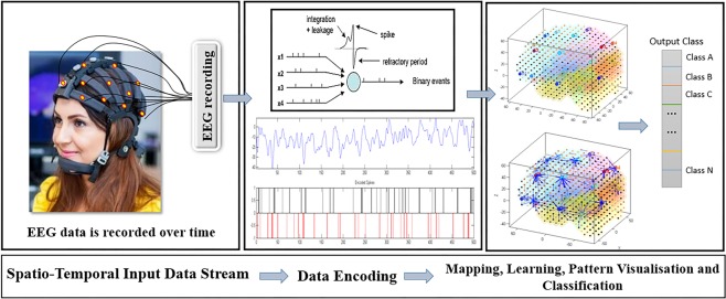
In today’s Research News article “Spiking Neural Network Modelling Approach Reveals How Mindfulness Training Rewires the Brain.” (See summary below or view the full text of the study at: https://www.ncbi.nlm.nih.gov/pmc/articles/PMC6478904/), Doborjeh and colleagues recruited adults and provided them with a 6-week, once a week for 90-120 minutes, mindfulness training. They separated the participants based upon a paper and pencil measure of depression into low depression, no depression, and high depression groups. Before and after training they measured the participants brain electrical activity with an electroencephalogram (EEG). They then employed a sophisticated data analysis algorithm to follow bursts of electrical activity through the brain (Spiking Neural Network).
They found that the no depression and low depression groups had overall higher activation than the high depression group. After mindfulness training the high depression group had higher activation at the frontal, temporal, frontocentral, and centroparietal sites, while the no depression group had higher activation of the frontal and occipitalparietal cortical areas, and the low depression group had higher activation at the frontal, temporal, and frontocentral sites.
The results are interesting and suggest that the Spiking Neural Network analysis of the electroencephalogram (EEG) can detect differences in brain activation in groups varying in levels of depression and can also detect neuroplastic changes resulting from mindfulness training. This is an important demonstration as it verifies that the easy, non-invasive, and economical EEG recording technique can be used to assess the details of neural function and the changes in neural function that may occur after an intervention.
Additionally, the results suggest that depressed individuals have quite low levels of brain activation which may, in part, be responsible for their depression. The results also show that mindfulness training in these depressed individuals can, to some extent, raise the levels of brain activation. This may be responsible, in part, for the ability of mindfulness training to decrease depression levels in depressed individuals. In addition, the results suggest that even in low and non-depressed individuals, mindfulness training can further increase brain activation. This may be responsible for the improvement in emotion regulation and mood in normal individuals that is produced by mindfulness training.
So, alter brain activation regardless of depression with mindfulness.
“There is emerging evidence that mindfulness meditation might cause neuroplastic changes in the structure and function of brain regions involved in regulation of attention, emotion and self-awareness.” – Britta Hőlzel
CMCS – Center for Mindfulness and Contemplative Studies
This and other Contemplative Studies posts are also available on Google+ https://plus.google.com/106784388191201299496/posts and on Twitter @MindfulResearch
Study Summary
Doborjeh, Z., Doborjeh, M., Taylor, T., Kasabov, N., Wang, G. Y., Siegert, R., & Sumich, A. (2019). Spiking Neural Network Modelling Approach Reveals How Mindfulness Training Rewires the Brain. Scientific reports, 9(1), 6367. doi:10.1038/s41598-019-42863-x
Abstract
There has been substantial interest in Mindfulness Training (MT) to understand how it can benefit healthy individuals as well as people with a broad range of health conditions. Research has begun to delineate associated changes in brain function. However, whether measures of brain function can be used to identify individuals who are more likely to respond to MT remains unclear. The present study applies a recently developed brain-inspired Spiking Neural Network (SNN) model to electroencephalography (EEG) data to provide novel insight into: i) brain function in depression; ii) the effect of MT on depressed and non-depressed individuals; and iii) neurobiological characteristics of depressed individuals who respond to mindfulness. Resting state EEG was recorded from before and after a 6 week MT programme in 18 participants. Based on self-report, 3 groups were formed: non-depressed (ND), depressed before but not after MT (responsive, D+) and depressed both before and after MT (unresponsive, D−). The proposed SNN, which utilises a standard brain-template, was used to model EEG data and assess connectivity, as indicated by activation levels across scalp regions (frontal, frontocentral, temporal, centroparietal and occipitoparietal), at baseline and follow-up. Results suggest an increase in activation following MT that was site-specific as a function of the group. Greater initial activation levels were seen in ND compared to depressed groups, and this difference was maintained at frontal and occipitoparietal regions following MT. At baseline, D+ had great activation than D−. Following MT, frontocentral and temporal activation reached ND levels in D+ but remained low in D−. Findings support the SNN approach in distinguishing brain states associated with depression and responsiveness to MT. The results also demonstrated that the SNN approach can be used to predict the effect of mindfulness on an individual basis before it is even applied.
https://www.ncbi.nlm.nih.gov/pmc/articles/PMC6478904/