Meditation Changes the Structures and Connectivity in the Brain
By John M. de Castro, Ph.D.
“[Meditation] practice appears to have an amazing variety of neurological benefits – from changes in grey matter volume to reduced activity in the “me” centers of the brain to enhanced connectivity between brain regions.” – Alice G. Walton
There has accumulated a large amount of research demonstrating that meditation practice has significant benefits for psychological, physical, and spiritual wellbeing. One way that meditation practices may produce these benefits is by altering the brain. The nervous system is a dynamic entity, constantly changing and adapting to the environment. It will change size, activity, and connectivity in response to experience. These changes in the brain are called neuroplasticity. Over the last decade neuroscience has been studying the effects of contemplative practices on the brain and has identified neuroplastic changes in widespread areas. In other words, meditation practice appears to mold and change the brain structures and connectivity, producing psychological, physical, and spiritual benefits, especially mindfulness.
In today’s Research News article “Meditation-induced effects on whole-brain structural and effective connectivity.” (See summary below or view the full text of the study at: https://www.ncbi.nlm.nih.gov/pmc/articles/PMC9232427/ ) De Filippi and colleagues recruited experienced meditators (> 1000 hours of experience) and control non-meditators and scanned their brains with functional Magnetic Resonance Imaging (fMRI) while at rest and while meditating.
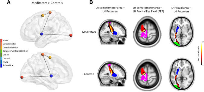
They found that there were significant differences in whole brain activity between meditation and at rest and that these differences differed between experienced meditators and controls. They also found that experienced meditators had increased connectivity between the left hemisphere sub networks including the somatomotor, dorsal attention, subcortical, and visual networks. Hence, they found neuroplastic changes in the brain produced by meditation practice.
“Now, as the popularity of mindfulness grows, brain imaging techniques are revealing that this ancient practice can profoundly change the way different regions of the brain communicate with each other – and therefore how we think – permanently.” – Tom Ireland
CMCS – Center for Mindfulness and Contemplative Studies
This and other Contemplative Studies posts are also available on Twitter @MindfulResearch
Study Summary
De Filippi, E., Escrichs, A., Càmara, E., Garrido, C., Marins, T., Sánchez-Fibla, M., Gilson, M., & Deco, G. (2022). Meditation-induced effects on whole-brain structural and effective connectivity. Brain structure & function, 227(6), 2087–2102. https://doi.org/10.1007/s00429-022-02496-9
Abstract
In the past decades, there has been a growing scientific interest in characterizing neural correlates of meditation training. Nonetheless, the mechanisms underlying meditation remain elusive. In the present work, we investigated meditation-related changes in functional dynamics and structural connectivity (SC). For this purpose, we scanned experienced meditators and control (naive) subjects using magnetic resonance imaging (MRI) to acquire structural and functional data during two conditions, resting-state and meditation (focused attention on breathing). In this way, we aimed to characterize and distinguish both short-term and long-term modifications in the brain’s structure and function. First, to analyze the fMRI data, we calculated whole-brain effective connectivity (EC) estimates, relying on a dynamical network model to replicate BOLD signals’ spatio-temporal structure, akin to functional connectivity (FC) with lagged correlations. We compared the estimated EC, FC, and SC links as features to train classifiers to predict behavioral conditions and group identity. Then, we performed a network-based analysis of anatomical connectivity. We demonstrated through a machine-learning approach that EC features were more informative than FC and SC solely. We showed that the most informative EC links that discriminated between meditators and controls involved several large-scale networks mainly within the left hemisphere. Moreover, we found that differences in the functional domain were reflected to a smaller extent in changes at the anatomical level as well. The network-based analysis of anatomical pathways revealed strengthened connectivity for meditators compared to controls between four areas in the left hemisphere belonging to the somatomotor, dorsal attention, subcortical and visual networks. Overall, the results of our whole-brain model-based approach revealed a mechanism underlying meditation by providing causal relationships at the structure-function level.
https://www.ncbi.nlm.nih.gov/pmc/articles/PMC9232427/