Change Your Brain’s Activity with Mindfulness
By John M. de Castro, Ph.D.
“The impact that mindfulness exerts on our brain is borne from routine: a slow, steady, and consistent reckoning of our realities, and the ability to take a step back, become more aware, more accepting, less judgmental, and less reactive. Just as playing the piano over and over again over time strengthens and supports brain networks involved with playing music, mindfulness over time can make the brain, and thus, us, more efficient regulators, with a penchant for pausing to respond to our worlds instead of mindlessly reacting.” – Jennifer Wolkin
There has accumulated a large amount of research demonstrating that mindfulness practices have significant benefits for psychological, physical, and spiritual wellbeing. Its positive effects are so widespread that it is difficult to find any other treatment of any kind with such broad beneficial effects on everything from mood and happiness to severe mental and physical illnesses. This raises the question of how meditation could do this. The nervous system is a dynamic entity, constantly changing and adapting to the environment. It will change size, activity, and connectivity in response to experience. These changes in the brain are called neuroplasticity. Over the last decade neuroscience has been studying the effects of contemplative practices on the brain and has identified neuroplastic changes in widespread areas. In other words, mindfulness practices appears to mold and change the brain, producing psychological, physical, and spiritual benefits.
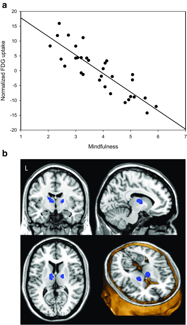
If mindfulness training can alter the nervous system then perhaps simply being a mindful individual will be associated with differences in the same brain regions. This idea was examined in today’s Research News article “Resting Brain Activity Related to Dispositional Mindfulness: a PET Study.” See summary below or view the full text of the study at: https://www.ncbi.nlm.nih.gov/pmc/articles/PMC5506209/, Gartenschläger and colleagues recruited normal and psychologically disturbed individuals and measured their levels of mindfulness, depression, and anxiety. The participants then underwent a brain scan for neural activity (Positron Emission Tomography, PET Scan).
They found that the higher the participant’s level of mindfulness, the lower the levels of both depression and anxiety. This is not surprising as mindfulness training has been shown repeatedly to produce lower levels of anxiety and depression. They also found that the higher the levels of mindfulness the higher the resting brain activity in the superior parietal lobule and in precuneus and superior parietal lobule and the lower the activity in the inferior frontal orbital gyrus and anterior thalamus.
These results are complex but the lower activity in the Thalamus may represent lower levels of general activation of the brain in mindful individuals. Also, the lower activity in the inferior frontal orbital gyrus may represent lower levels of language processing in mindful individuals, possibly indicating less internal language, thinking, with individuals high in mindfulness. In addition, the higher activity in the parietal lobe and precuneus may represent greater activity in the Default Mode Network (DMN) of which these structures are a part. The DMN is associated with a sense of self, self-referential thinking, and mind wandering. This suggests that mindful individuals while at rest, with their eyes closed, may be less activated (more at rest), have less internal language (thought), and have their minds wandering.
It may seem counterintuitive that mindful individuals’ minds may be wandering more as mindfulness has been shown to be associated with less mind wandering. But, the situation of lying in a scanner with eyes closed may be one in which discursive thought is perfectly appropriate. In any case, these are interesting results that add to our understanding of the brain systems involved in mindfulness. It will require considerable future research to paint a complete picture of the neural systems underlying mindfulness and being altered by mindfulness training.
So, change your brain’s activity with mindfulness.
“The practice of mindfulness can train our brains to have a new default. Instead of automatically falling into the stream of past or future rumination that ignites the depression loop, mindfulness draws our attention to the present moment. As we practice mindfulness, we actually start wiring neurons that balance the brain in a way that is naturally an antidepressant.” – Alex Korb
CMCS – Center for Mindfulness and Contemplative Studies
This and other Contemplative Studies posts are also available on Google+ https://plus.google.com/106784388191201299496/posts and on Twitter @MindfulResearch
Study Summary
Gartenschläger, M., Schreckenberger, M., Buchholz, H.-G., Reiner, I., Beutel, M. E., Adler, J., & Michal, M. (2017). Resting Brain Activity Related to Dispositional Mindfulness: a PET Study. Mindfulness, 8(4), 1009–1017. http://doi.org/10.1007/s12671-017-0677-2
Abstract
Mindfulness denotes a state of consciousness characterized by receptive attention to and awareness of present events and experiences. As a personality trait, it constitutes the ability to become aware of mental activities such as sensations, images, feelings, and thoughts, and to disengage from judgment, conditioned emotions, and their cognitive processing or automatic inhibition. Default brain activity reflects the stream of consciousness and sense of self at rest. Analysis of brain activity at rest in persons with mindfulness propensity may help to elucidate the neurophysiological basis of this important mental trait. The sample consisted of 32 persons—23 with mental disorders and 9 healthy controls. Dispositional mindfulness (DM) was operationalized by Mindful Attention Awareness Scale (MAAS). Brain activity at rest with eyes closed was assessed by fluorodeoxyglucose positron emission tomography (F-18-FDG PET). After adjustment for depression, anxiety, age and years of education, resting glucose metabolism in superior parietal lobule and left precuneus/Brodmann area (BA) 7 was positively associated with DM. Activity of the left inferior frontal orbital gyrus (BA 47) and bilateral anterior thalamus were inversely associated with DM. DM appears to be associated with increased metabolic activity in some core area of the default mode network (DMN) and areas connected to the DMN, such as BA 7, hosting sense of self functions. Hypometabolism on the other hand was found in some nodes connected to the DMN, such as left inferior frontal orbital gyrus and bilateral thalamus, commonly related to functions of memory retrieval, decision making, or outward attention.